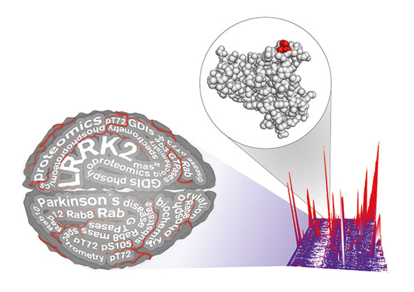
Parkinson’s researchers used proteomics to identify Rab proteins as a physiological substrate of LRRK2, a Parkinson’s drug target. This finding may accelerate current research and open a novel therapeutic avenue. Credit: MPI f. Biochemistry
An international team has discovered that the LRRK2 kinase regulates cellular trafficking by deactivating Rab proteins. This finding illuminates a novel route for therapeutic development and may accelerate testing of LRRK2 inhibitors as a disease-modifying therapy for Parkinson’s, the second most common neurodegenerative disease.
An international public-private research consortium has identified and validated a cellular role of a primary Parkinson’s disease drug target, the LRRK2 kinase. This important finding, to be published in the online, open-access eLife journal, illuminates a novel route for therapeutic development and intervention testing for Parkinson’s, the second most common neurodegenerative disease after Alzheimer’s.
A team of investigators from the Max Planck Institute of Biochemistry, the University of Dundee, The Michael J. Fox Foundation for Parkinson’s Research (MJFF), GlaxoSmithKline (GSK), and MSD contributed unique tools and expertise toward rigorous systematic testing that determined the LRRK2 kinase regulates cellular trafficking by deactivating certain Rab proteins (3, 8, 10 and 12).
Mutations in the LRRK2 gene are the greatest known genetic contributor to Parkinson’s disease. Pharmaceutical companies are developing LRRK2 kinase inhibitors to correct the effects of those mutations and treat Parkinson’s disease. This new breakthrough finding that links mutant LRRK2 to inappropriate deactivation of Rab function unlocks more than 20 years of accumulated knowledge of the roles of Rab proteins. This knowledge can now be integrated to significantly improve our understanding of LRRK2 dysfunction in the Parkinson’s disease process.
“The pathological cascade leading to brain diseases such as Parkinson’s likely includes many cellular players,” said Matthias Mann, Ph.D., Director of the Department of Proteomics and Signal Transduction at the Max Planck Institute of Biochemistry. “The identification of this LRRK2 substrate gives us a central piece in this puzzle and another potential place to intervene in the disease process.”
Marco Baptista, Ph.D., MJFF Senior Associate Director of Research Programs, said, “Identification of Rab proteins as a LRRK2 substrate presents a tool to measure the impact of these inhibitors not only on LRRK2 levels but also on LRRK2 function. This critical component will advance the development of these therapies to slow or stop Parkinson’s disease, patients’ greatest unmet need.”
This MJFF-led consortium used a combination of tools in the discovery, including a knock-in mouse model of the most common LRRK2 mutation strongly associated with Parkinson’s (created by GSK), a second knock-in LRRK2 mouse model generated by MJFF, LRRK2 kinase inhibitors from GSK and Merck, and state-of-the-art mass spectrometry. These tools — and the collaborative spirit that united the partners — were necessary to make this finding.
“This unique model of collaboration and our systematic approach across laboratories using advanced technologies and layers of confirmation provide a firm foundation from which to continue this line of investigation and further refine our understanding of the LRRK2 Rab relationship,” said Dario Alessi, Ph.D., Director of the Protein Phosphorylation and Ubiquitylation Unit at the University of Dundee.
With additional MJFF funding, this research group now is working to further characterize the Rab proteins modified by LRRK2 and to understand how an imbalance in cellular trafficking leads to the degeneration of neurons seen in Parkinson’s disease.
Reference: “Phosphoproteomics reveals that Parkinson’s disease kinase LRRK2 regulates a subset of Rab GTPases” by M. Steger, F. Tonelli, G. Ito, P. Davies, M. Trost, M. Vetter, S. Wachter, E. Lorentzen, G. Duddy, S. Wilson, M. A. S. Baptista, B. K. Fiske, M. J. Fell, J. A. Morrow, A. D. Reith, D. R. Alessi and M. Mann,28 January 2016, eLife.
DOI: 10.7554/eLife.12813


Parkinson’s disease is usually treated by giving medicine Levodopa. But excessive dopamine conversion by CNS is curtailed by giving another tablet D-Dopa to neutralise the effect. Whereas L-Dopa can cross blood brain barrier whereas Dopamine cannot trickle out from the brain to the CNS system. This article suggests another type of inhibitor , but this time LRRK2 kinase regulation so that the essential dopamine penetrates through the cells to produce the maximum effect for Parkinson’s disease. The degeneration of Parkinson’s disease further can be only delayed however more than the current understanding. After all the brain cells has stopped the production of Dopamine which is supplemental from the tablets which undergoes decomposition to Dopamine. The original stoppage of natural dopamine causes has not been addressed yet. Thank You.