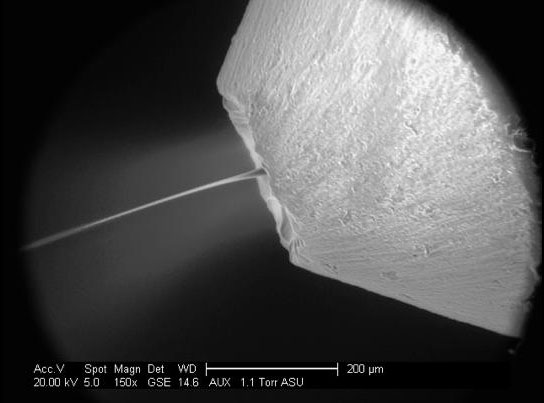
Scientists embedded tiny protein crystals in an oily solution that mimics the supportive environment of the cell membrane, and then squirted them through a micro-jet into the path of a powerful X-ray laser. By analyzing the diffraction patterns made by X-rays scattering off the crystals, they were able to determine the protein’s structure. A key challenge was adjusting the viscosity of the oily solution, called a “lipidic sponge phase,” so it wouldn’t clog the micro-jet’s nozzle, shown here. Credit: Richard Neutze
New research at SLAC’s Linac Coherent Light Source (LCLS) has shown a promising new way to collect data on membrane proteins in the human body. The method involves embedding tiny protein crystals in an oily paste and then hitting them with a powerful X-ray laser to determine the protein’s structure.
Many membrane proteins serve as gateways in and out of the cell. Because they act as “traffic control” for infectious agents and disease-fighting drugs, they are the targets of more than 60 percent of all drugs on the market. Yet of the estimated 30,000 membrane proteins in the human body, scientists understand the detailed structures of only 18.
Now experiments at SLAC’s Linac Coherent Light Source (LCLS) have shown a promising new way to collect data on these elusive proteins. Researchers embedded tiny protein crystals in an oily paste that mimics the supportive environment of the cell membrane, and then hit them with a powerful X-ray laser to determine the protein’s structure. They reported their results in the March issue of Nature Methods.
Until now, drug design based on membrane protein structures has been difficult; it simply took too much time and money to crystallize and probe the finicky proteins, which become unstable when taken out of their lipid environment, said Richard Neutze of the University of Gothenburg in Sweden, a co-author of the study. “The chances of failure are so high,” he said, adding that the new method “could help us increase knowledge about how the body functions and who we are as humans.”
Earlier studies at the LCLS determined that tiny protein crystals could be used to analyze the rough molecular structure of an important membrane protein complex called Photosystem I. Since these small crystals are much easier to prepare than the big ones required by traditional methods, the technique opened the door to understanding tens of thousands of proteins that were previously out of reach. Scientists squirted a solution containing the tiny crystals into the path of the X-ray laser beam. X-rays scattering off electrons in the protein’s atoms formed diffraction patterns that could be used to determine the protein structure.
Researchers have also achieved good results with very challenging membrane proteins by embedding them in a fatty paste before crystallization.
Scientists wondered whether that approach would work for experiments at the LCLS. Since the nozzle that squirts samples into the path of the X-ray laser has a very small opening, getting the viscosity of the paste just right would be a challenge: If it’s too thick, it will not form a thin enough jet, and may even clog the nozzle.
In this experiment, researchers mixed the membrane protein – a photosynthetic reaction center found in some bacteria – with a lipid called monoolein that had been thinned with water, and then added crystallization agents. After filtering out the largest crystals, they injected the liquid suspension, known as a “lipidic sponge phase,” through a micro-jet across the X-ray laser beam. They recorded 365,035 images in less than two hours, and analyzed 265 diffraction patterns to determine the protein’s structure.
This particular experiment was carried out on a protein whose structure was already known in great detail. It mapped the protein with a resolution of 8.2 Angstroms, compared to less than 2 Angstroms in previous, high-resolution studies. Nevertheless, it demonstrates that the method works and sets the stage for more detailed studies at the LCLS.
LCLS has already made available shorter wavelengths of X-rays and doubled the rate at which X-ray laser pulses can hit samples, from 60 to 120 pulses per second. Neutze said he expects these developments will soon make it possible to get high-resolution structures for membrane proteins, giving researchers a powerful tool for understanding at a molecular level why people become ill and what can be done to treat them. The work also has implications for energy research, since scientists are trying to reverse-engineer the photosynthetic processes that plants use to harvest energy from the sun, which takes place in membrane protein complexes.
The team’s next goal is to collect the approximately 10,000 diffraction images they estimate are needed to make high-resolution maps of protein structure. This first step in successfully collecting 265 images bodes well for future applications, the paper concludes.
Reference: “Lipidic phase membrane protein serial femtosecond crystallography” by Linda C Johansson, David Arnlund, Thomas A White, Gergely Katona, Daniel P DePonte, Uwe Weierstall, R Bruce Doak, Robert L Shoeman, Lukas Lomb, Erik Malmerberg, Jan Davidsson, Karol Nass, Mengning Liang, Jakob Andreasson, Andrew Aquila, Saša Bajt, Miriam Barthelmess, Anton Barty, Michael J Bogan, Christoph Bostedt, John D Bozek, Carl Caleman, Ryan Coffee, Nicola Coppola, Tomas Ekeberg, Sascha W Epp, Benjamin Erk, Holger Fleckenstein, Lutz Foucar, Heinz Graafsma, Lars Gumprecht, Janos Hajdu, Christina Y Hampton, Robert Hartmann, Andreas Hartmann, Günter Hauser, Helmut Hirsemann, Peter Holl, Mark S Hunter, Stephan Kassemeyer, Nils Kimmel, Richard A Kirian, Filipe R N C Maia, Stefano Marchesini, Andrew V Martin, Christian Reich, Daniel Rolles, Benedikt Rudek, Artem Rudenko, Ilme Schlichting, Joachim Schulz, M Marvin Seibert, Raymond G Sierra, Heike Soltau, Dmitri Starodub, Francesco Stellato, Stephan Stern, Lothar Strüder, Nicusor Timneanu, Joachim Ullrich, Weixiao Y Wahlgren, Xiaoyu Wang, Georg Weidenspointner, Cornelia Wunderer, Petra Fromme, Henry N Chapman, John C H Spence and Richard Neutze, 29 January 2012, Nature Methods.
DOI: 10.1038/nmeth.1867
Members of the large international research team represented more than a dozen institutions, including the Linac Coherent Light Source and Photon Ultrafast Laser Science and Engineering Center at SLAC; Gothenburg and Uppsala universities in Sweden; Arizona State University; the Center for Free-Electron Laser Science at DESY in Hamburg, Germany; DESY; the University of Hamburg; the Max Planck institutes for nuclear physics, extraterrestrial physics, semiconductor laboratory (Halbleiterlabor) and medical research; and the Advanced Light Source at the DOE’s Berkeley Lab.


Be the first to comment on "Examining Membrane Proteins by X-Ray Laser"