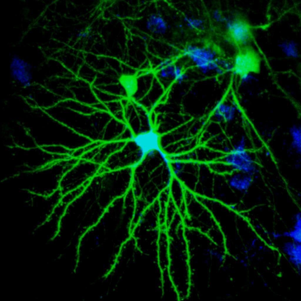
Neurons (nerve cells) are labeled with green fluorescent protein, and other neurons in the brain are labeled in the background with either red or blue tracers. The small bulbs (i.e., dendritic spines) on the spidery dendrites show places where nerve cells connect and communicate, called synapses, and when these spines shrank over time, this predicted vocal degradation in the songbirds. Credit: Katie Tschida, Duke Department of Neurobiology
It appears that deafness penetrates much more rapidly and deeply into the brain than previously thought. While studying songbirds, neurologists discovered that when their hearing was lost, their songs fell apart much like human vocalization does, making them an ideal subject to study how hearing loss may affect the parts of the brain that control vocalization.
Portions of a songbird’s brain that control how it sings have been shown to decay within 24 hours of the animal losing its hearing.
The findings, by researchers at Duke University Medical Center, show that deafness penetrates much more rapidly and deeply into the brain than previously thought. As the size and strength of nerve cell connections visibly changed under a microscope, researchers could even predict which songbirds would have worse songs in coming days.
“When hearing was lost, we saw rapid changes in motor areas in that control song, the bird’s equivalent of speech,” said senior author Richard Mooney, PhD, professor of neurobiology at Duke. “This study provided a laser-like focus on what happens in the living songbird brain, narrowed down to the particular cell type involved.”
The study was published in Neuron journal online on March 8, 2012.
Like humans, songbirds depend on hearing to learn their mating songs – males that sing poorly don’t attract mates, so hearing a song, learning it, and singing correctly are all critical for songbird survival. Songbirds also resemble humans and differ from most other animals in that their songs fall apart when they lose their hearing, and this feature makes them an ideal organism to study how hearing loss may affect the parts of the brain that control vocalization, Mooney said.

Top – Spectrogram of a song, before songbird became deaf. Bottom – Spectrogram of a song, after same songbird became deaf. This bird (a male, because only male zebra finches sing) was around 90 days post-hatch when he became deaf. This is around the age at which zebra finches sexually mature, so he is considered a young adult. Two weeks after deafening, this song sounds messier, and literally looks more degraded on these representations of the song notes.
“I will go out on a limb and say that I think similar changes also occur in human brains after hearing loss, specifically in Broca’s area, a part of the human brain that plays an important role in generating speech and that also receives inputs from the auditory system,” Mooney said.
About 30 million Americans are hard of hearing or deaf. This study could shed light on why and how some people’s speech changes as their hearing starts to decline, Mooney said.
“Our vocal system depends on the auditory system to create intelligible speech. When people suffer profound hearing loss, their speech often becomes hoarse, garbled, and harder to understand, so not only do they have trouble hearing, they often can’t speak fluently any more,” Mooney said.
The nerve cells that showed changes after deafening send signals to the basal ganglia, a part of the brain that plays a role in learning and initiating motor sequences, including the complex vocal sequences that make up birdsong and speech.
Although other studies had looked at the effects of deafening on neurons in auditory brain areas, this is the first time that scientists have been able to watch how deafening affects connections between nerve cells in a vocal motor area of the brain in a living animal, said Katie Tschida, PhD, a postdoctoral research associate in the Mooney laboratory who led the study.
Using a protein isolated from jellyfish that can make songbird nerve cells glow bright green when viewed under a laser-powered microscope, they were able to determine that deafening triggered rapid changes to the tiny connections between nerve cells, called synapses, which are only one-thousandth of a millimeter across.
“I was very surprised that the weakening of connections between nerve cells was visible and emerged so rapidly — over the course of days these changes allowed us to predict which birds’ songs would fall apart most dramatically,” Tschida said. “Considering that we were only tracking a handful of neurons in each bird, I never thought we’d get information specific enough to predict such a thing.”
Reference: “Deafening Drives Cell-Type-Specific Changes to Dendritic Spines in a Sensorimotor Nucleus Important to Learned Vocalizations” by Katherine A. Tschida and Richard Mooney, 8 March 2012, Neuron.
DOI: 10.1016/j.neuron.2011.12.038
The research was supported by the National Science Foundation and the National Institute on Deafness and Other Communication Disorders.


Be the first to comment on "Hearing Loss Rapidly Changes Brain Motor Areas That Control Song"