
Neuroscientists find they can predict how well someone can see based on the unique structure of their primary visual cortex.
Visual Cortex Size, Brain Tissue Can Predict How Well We See
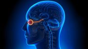
In many ways, the eye functions as a camera, with the retina acting as the photographic film (or CCD in a digital camera.) However, you don’t actually see anything without your brain, which receives the visual signals from the eye via the optic nerve.
The size of our primary visual cortex and the amount of brain tissue we have dedicated to processing visual information at certain locations of visual space can predict how well we can see, a team of neuroscientists has discovered. Its study, which appears today (June 13, 2022) in the journal Nature Communications, reveals a new link between brain structure and behavior.
“We have found that we can predict how well someone can see based on the unique structure of their primary visual cortex,” explains lead author Marc Himmelberg, a postdoctoral researcher in New York University’s Center for Neural Science and Department of Psychology. “By showing that individual variation in the structure of the human visual brain is linked to variation in visual functioning, we can better understand what underlies differences in how people perceive and interact with their visual environment.”
As with fingerprints, the bumps and grooves on each person’s brain surface are unique. However, the significance of these differences is not fully understood, especially when it comes to their impact on behavior, such as distinctions in our ability to see.
In the study published in Nature Communications, Himmelberg and his co-authors, Jonathan Winawer and Marisa Carrasco, professors in NYU’s Center for Neural Science and Department of Psychology, sought to illuminate the relevance of these brain traits to how we see.
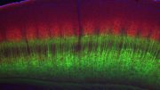
The primary visual cortex (V1) is arranged into a map of the image projected from the eye. But like many kinds of maps, it is distorted, with some parts of the image enlarged compared to others.
“Think of a subway map of New York City which makes Staten Island look smaller than Manhattan,” explains Winawer. “The map maintains some degree of accuracy, but it enlarges regions likely to be of broader interest. Similarly, V1 enlarges the center of the image we see—that is, where our eyes are fixating—relative to the periphery.”
This is because V1 has more tissue dedicated to the center of our field of view. Likewise, V1 also enlarges locations to the left and right of where our eyes are fixating relative to locations above or below, again because of differences in the arrangement of cortical tissue.
Using functional magnetic resonance imaging (fMRI), the scientists mapped the primary visual cortex (or “V1”) size of more than two dozen humans. The researchers also measured the quantity of V1 tissue these individuals have dedicated to processing visual information from different locations in their field of view—locations to the left, right, above, and below fixation.
These participants also undertook a task designed to assess the quality of their vision at the same locations in their field of view as the V1 measurements. The participants discriminated among the orientation of patterns shown on a computer screen, which were used to gauge “contrast sensitivity,” or the ability to make distinctions among images.
Their results showed that differences in V1 surface area could predict measurements of people’s contrast sensitivity. First, people with a large V1 had better overall contrast sensitivity than did those with a small V1 (the largest surface area being 1,776 square millimeters [mm2] and the smallest being 832 mm2). Second, people whose V1 had more cortical tissue processing visual information from a specific region in their field of view had higher contrast sensitivity at that region relative to those with less cortical tissue dedicated to the same region. Third, across participants, higher contrast sensitivity at a specific location (e.g., left) than at another location equidistant from fixation (e.g., above) corresponded to regions with more or less cortical tissue, respectively.
“In sum, the more local V1 surface area dedicated to encoding a specific location, the better the vision at that location,” concludes Carrasco. “Our findings show differences in visual perception are inextricably linked to differences in the structure of the primary visual cortex in the brain.”
Reference: “Linking individual differences in human primary visual cortex to contrast sensitivity around the visual field” by Marc M. Himmelberg, Jonathan Winawer and Marisa Carrasco, 13 June 2022, Nature Communications.
DOI: 10.1038/s41467-022-31041-9
The research was supported by a grant from the National Institutes of Health (R01-EY027401).


Wow , super interesting.
Amazing subject , expecting more info
Great article and so easy to understand. Thanks!
So saying it with out saying it.double speak. better Vision equals smarter.just what kinds of smart and how much smarter are we talking here compared to what the blind?
Can we improve or vision?
cortical area ∝ vision quality – does it even need such a test ?