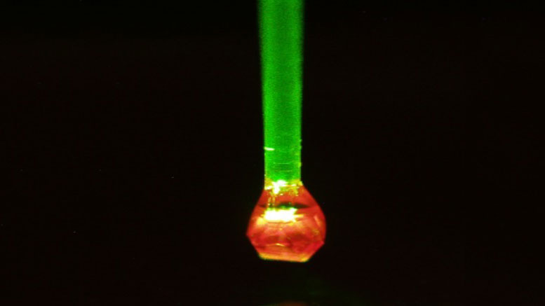
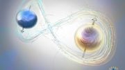
Green laser light transmitted via an optical fiber excites nitrogen atoms in a diamond, causing it to fluoresce with a red light. The brightness of a nitrogen atom at the edge of the diamond lattice allows conclusions to be drawn about the magnetic signals from a sample on the surface of the sensor. University of Stuttgart
A team of researchers has developed a quantum sensor that makes it possible to use nuclear magnetic resonance scanning to investigate the structure of individual proteins atom by atom.
Nuclear magnetic resonance scanners, as are familiar from hospitals, are now extremely sensitive. A quantum sensor developed by a team headed by Professor Jörg Wrachtrup at the University of Stuttgart and researchers at the Max Planck Institute for Solid State Research in Stuttgart, now makes it possible to use nuclear magnetic resonance scanning to even investigate the structure of individual proteins atom by atom. In the future, the method could help to diagnose diseases at an early stage by detecting the first defective proteins.
Many diseases have their origins in defective proteins. As proteins are important biochemical motors, defects can lead to disturbances in metabolism. Defective prions, which cause brain damage in BSE and Creutzfeldt-Jakob disease, are one example. Pathologically changed prions have defects in their complex molecular structure. The problem: individual defective proteins can likewise induce defects in neighboring intact proteins via a sort of domino effect and thus trigger a disease. It would therefore be very useful if doctors could detect the first, still individual prions with the wrong structure. It has, however, not been possible to date to elucidate the structure of one individual biomolecule.
In an article published in “Science”, a team of researchers from Stuttgart has now presented a method that can be used in the future for the reliable investigation of individual biomolecules. This is important not only for fighting diseases, but also for chemical and biochemical basic research.
The method involves the miniaturization as it were of the nuclear magnetic resonance tomography (NMR) known from medical engineering, which is usually called MRI scanning in the medical field. NMR makes use of a special property of the atoms – their spin. In simple terms, spin can be thought of as the rotation of atomic nuclei and electrons about their own axis, turning the particles into tiny, spinning bar magnets. How these magnets behave is characteristic for each type of atom and each chemical element. Each particle thus oscillates with a specific frequency.
In medical applications, it is normal for only one type of atom to be detected in the body – hydrogen, for example. The hydrogen content in the different tissues allows the interior of the body to be distinguished with the aid of various contrasts.
Structural resolution at the atomic level
When elucidating the structure of biomolecules, on the other hand, each individual atom must be determined and the structure of the biomolecule then deciphered piece by piece. The crucial aspect here is that the NMR detectors are so small that they achieve nanometer-scale resolution and are so sensitive that they can measure individual molecules exactly. It is more than four years ago that the researchers working with Jörg Wrachtrup first designed such a small NMR sensor; it did not, however, allow them to distinguish between individual atoms.
To achieve atomic-level resolution, the researchers must be able to distinguish between the frequency signals they receive from the individual atoms of a molecule – in the same way as a radio identifies a radio station by means of its characteristic frequency. The frequencies of the signals emitted by the atoms of a protein are those frequencies at which the atomic bar magnets in the protein spin. These frequencies are very close together, as if the transmission frequencies of radio stations all tried to squeeze themselves into a very narrow bandwidth. This is the first time the researchers in Stuttgart have achieved a frequency resolution at which they can distinguish individual types of atoms.
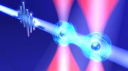
“We have developed the first quantum sensor that can detect the frequencies of different atoms with sufficient precision and thus resolve a molecule almost into its individual atoms,” says Jörg Wrachtrup. It is thus now possible to scan a large biomolecule, as it were. The sensor, which acts as a minute NMR antenna, is a diamond with a nitrogen atom embedded into its carbon lattice close to the surface of the crystal. The physicists call the site of the nitrogen atom the NV center: N for nitrogen and V for vacancy, which refers to a missing electron in the diamond lattice directly adjacent to the nitrogen atom. Such an NV center detects the nuclear spin of atoms located close to this NV center.
Simple yet very precise
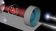
The spin frequency of the magnetic moment of an atom that has just been measured is transferred to the magnetic moment in the NV center, which can be seen with a special optical microscope as a change in color.
The quantum sensor achieves such high sensitivity, as it can store frequency signals of an atom. One single measurement of the frequency of an atom would be too weak for the quantum sensor and possibly too noisy. The memory allows the sensor to store many frequency signals over a longer period of time, however, and thus tune itself very precisely to the oscillation frequency of an atom – in the same way as a high-quality short-wave receiver can clearly resolve radio channels that are very close to each other.
This technology has other advantages apart from its high resolution: it operates at room temperature and, unlike other high-sensitivity NMR methods used in biochemical research, it does not require a vacuum. Moreover, these other methods generally operate close to absolute zero – minus 273.16 degrees Celsius – necessitating complex cooling with helium.
Future field of application: brain research
Jörg Wrachtrup sees not one but several future fields of application for his high-resolution quantum sensors. “It is conceivable that, in the future, it will be possible to detect individual proteins that have undergone a noticeable change in the early stage of a disease and which have so far been overlooked.” Furthermore, Wrachtrup is collaborating with an industrial company on a slightly larger quantum sensor that could be used in the future to detect the weak magnetic fields of the brain. “We call this sensor the brain reader. We hope it will help us to decipher how the brain works – and it would be a good complement to the conventional electrical devices derived from the EEG” – the electroencephalogram. For the brain reader, Wrachtrup is already working with his industrial partner on a holder and a casing so that the device is easy to wear and operate on a day-to-day basis. To reach this point, however, it will take at least another ten years of research.
Reference: “Nanoscale nuclear magnetic resonance with chemical resolution” by Nabeel Aslam, Matthias Pfender, Philipp Neumann, Rolf Reuter, Andrea Zappe, Felipe Fávaro De Oliveira, Andrej Denisenko, Hitoshi Sumiya, Shinobu Onoda, Junichi Isoya and Jörg Wrachtrup, 1 June 2017, Science.
DOI: 10.1126/science.aam8697


Be the first to comment on "Researchers Develop a Nuclear Magnetic Resonance Scanner for Individual Proteins"