
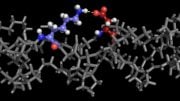
Catarina Moura’s ‘Christmas tree’ is comprised of laser-based images of collagen and fat cells.
At first glance, a pair of award-winning images created by University of Southampton postgraduate researcher Catarina Moura seem to have a seasonal theme. But look more closely and you’ll see that the component parts of the pictures (or micrographs) of a Christmas tree and seasonal wreath are actually comprised of stem cells created using innovative laser-based imaging techniques used as part of a regenerative medicine research program in Southampton.
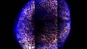
The green circular objects in the pictures are collagen cells and the red ‘dots’ are fat cells, both extracted from human bone marrow which Catarina has color-enhanced electronically to bring out their detail. Normally, the collagen cells that appear green on Catarina’s tree and wreath images would be bluer in color but, she says, the fat cells would certainly appear red with the lasers used.
“I’ve chosen green for the collagen fibers in my images because when you use the labeling technique typically you use a stain that fluoresces as green and, because a scientist would usually relate to that green color when looking at labeled collagen fiber, I decided to use the same color and create something more festive at the same time,” says Catarina.
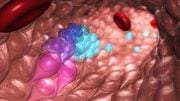
Working with Skeletal Biologists at Southampton General Hospital, Catarina is investigating new optical techniques to monitor the development of the cells, used in new regenerative medicine approaches – in this case, to create and grow cartilage from human stem cells. Her Ph.D. is focused on developing a novel label-free imaging approach for assessing human stem cells and skeletal regeneration non-destructively and non-invasively.

Micrograph of a Christmas wreath comprised of stem cells. Catarina Moura, University of Southampton
“I’m working with Professor Richard Oreffo and Dr. Rahul Tare from the University’s Center for Human Development, Stem Cells and Regeneration who are trying to create and grow cartilage in the lab using a patient’s own (autologous) stem cells to then be implanted back into the patient if they have a cartilage defect,” she explains. “My part of the project is to develop and use techniques to make it easier to monitor the development of the cells into cartilage in real time which is important to knowing if and when you can use it for the patient. If it’s successful, you can use the same cartilage to create the new tissue so it’s very important for us to get the monitoring right.”
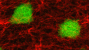
Traditional techniques involve labeling or injecting the cells with stains or fluorophores – fluorescent compounds that ‘glow’ when exposed to light – to detect their intricate structures. Under the tutelage of her Ph.D. supervisor, Southampton’s Sumeet Mahajan, Professor in Molecular Biophotonics & Imaging in Chemistry & Institute for Life Sciences (IfLS), Catarina is using ultra-fast lasers to achieve the same effect but in a less invasive way.
“Traditional techniques to detect whether the cartilage is developing can be disruptive and, in many cases, destructive,” Catarina explains. “Our process has not been used before. What we are trying to do is introduce to biology techniques normally used in chemistry or physics, using inherent chemical or structural properties of the human stem cells. Currently, for validation we still need to do the standard exercises alongside our new techniques to be able to compare the two sets of results and, of course, using ultra-fast lasers we need to ensure that everything is optimized before it can go to the clinic, especially the exposure time.
“The massive advantage with our stain-less laser-based imaging approaches is that you can use the stem cell sample without having to interrupt the developmental process in real time, you don’t need to perform any cell disruption and there is no photobleaching (fading) which is fairly common with fluorescent material,” Catarina enthused. “Just put the bioengineered cartilage under the microscope and you have the image.”
Professor Richard Oreffo added: “Crucially, unlike current standard staining-based methods the stain-less imaging approach is translatable to the clinic as the stem cells are not harmed or disrupted in any way. Hence, the technology can be used to objectively assess development and screen stem cells to be absolutely sure before using them for therapy.”
Professor Mahajan, concluded: “This work perfectly exemplifies highly exciting cross-faculty interdisciplinary research that is pushing boundaries to achieve high impact. Ph.D. funding by the IfLS for Catarina kick-started the collaboration between Richard and Rahul at the Institute for Developmental Sciences and us, which otherwise might have been difficult, that has led to exciting results, some stunning images and insight that has the potential to change people’s lives using stem cell therapy.”


Be the first to comment on "Seasonal Color-Enhanced Images Reveal the Science Behind Stem Cells"