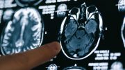
Investigators have uncovered brain changes in patients up to six months after they recovered from COVID-19.
Scientists uncovered brain changes in patients up to six months after they recovered from COVID-19 by using a special type of MRI. This is according to a study that will be presented at the annual meeting of the Radiological Society of North America (RSNA) next week.
According to the U.S. Centers for Disease Control and Prevention (CDC), approximately one in five adults will develop long-term effects from COVID-19. Difficulty thinking or concentrating, sleep problems, headache, lightheadedness, change in smell or taste, pins-and-needles sensation, and depression or anxiety are all neurological symptoms associated with long COVID. However, research studies have found that COVID-19 may be associated with changes to the heart, lungs, or other organs even in asymptomatic patients.
As more people become infected and recover from COVID-19, research has begun to emerge, focusing on the lasting consequences of the disease. These are known as post-COVID conditions, which are also known by a myriad of names including long COVID, long-haul COVID, post-acute COVID-19, post-acute sequelae of SARS CoV-2 infection (PASC), long-term effects of COVID, and chronic COVID.
For this study, researchers used susceptibility-weighted imaging to analyze the effects that COVID-19 has on the brain. Magnetic susceptibility denotes how much certain materials, such as blood, iron, and calcium, will become magnetized in an applied magnetic field. This ability aids in the detection and monitoring of a host of neurologic conditions including microbleeds, vascular malformations, brain tumors, and stroke.
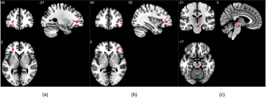
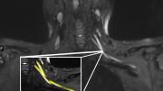
Group analysis on susceptibility-weighted imaging exhibiting higher susceptibility-weighted imaging values in the COVID group when compared to healthy controls. Three significant clusters were found primarily in the white matter regions of the pre-frontal cortex and in the brainstem. The clusters (a) and (b) are observed bilaterally in the cerebral white matter near the orbitofrontal gyrus, whereas (c) lies in the midbrain region. Credit: RSNA and Sapna S. Mishra
“Group-level studies have not previously focused on COVID-19 changes in magnetic susceptibility of the brain despite several case reports signaling such abnormalities,” said study co-author Sapna S. Mishra, a Ph.D. candidate at the Indian Institute of Technology in Delhi. “Our study highlights this new aspect of the neurological effects of COVID-19 and reports significant abnormalities in COVID survivors.”
The researchers analyzed the susceptibility-weighted imaging data of 46 COVID-recovered patients and 30 healthy controls. Imaging was done within six months of recovery. Among patients with long COVID, the most commonly reported symptoms were fatigue, trouble sleeping, lack of attention, and memory issues.
“Changes in susceptibility values of brain regions may be indicative of local compositional changes,” Mishra said. “Susceptibilities may reflect the presence of abnormal quantities of paramagnetic compounds, whereas lower susceptibility could be caused by abnormalities like calcification or lack of paramagnetic molecules containing iron.”
MRI results showed that patients who recovered from COVID-19 had significantly higher susceptibility values in the frontal lobe and brain stem compared to healthy controls. The clusters obtained in the frontal lobe primarily show differences in the white matter.
“These brain regions are linked with fatigue, insomnia, anxiety, depression, headaches, and cognitive problems,” Mishra said.
Portions of the left orbital-inferior frontal gyrus (a key region for language comprehension and production) and right orbital-inferior frontal gyrus (associated with various cognitive functions including attention, motor inhibition, and imagery, as well as social cognitive processes) and the adjacent white matter areas made up the frontal lobe clusters.
The researchers also found a significant difference in the right ventral diencephalon region of the brain stem. This region is associated with many crucial bodily functions, including coordinating with the endocrine system to release hormones, relaying sensory and motor signals to the cerebral cortex and regulating circadian rhythms (the sleep-wake cycle).
“This study points to serious long-term complications that may be caused by the coronavirus, even months after recovery from the infection,” Mishra said. “The present findings are from the small temporal window. However, the longitudinal time points across a couple of years will elucidate if there exists any permanent change.”
The researchers are conducting a longitudinal study on the same patient cohort to determine whether these brain abnormalities persist over a longer time frame.
Co-authors are Rakibul Hafiz, Ph.D., Tapan Gandhi, Ph.D., Vidur Mahajan, M.B.B.S., Alok Prasad, M.D., and Bharat Biswal, Ph.D.
Meeting: 108th Scientific Assembly and Annual Meeting of the Radiological Society of North America

It would be informative for average person to know “IF” the pst affects are fir original Covid-19 virus or the later less virulent versions-if ant difference.
I have too many friends that were vaccinated and boosted and boosted and still got Covid later variants.
Now we hear research and reports of cardiac problems with vaccinated persons under 50 years of age. Indicating that the Covid shots are not thoroughly vetted as is a real vaccine. Remember the Covid shots were released under emergency measures and not tested as thoroughly as standard practice due to the spread of the Chinese virus
You got me until I read those last two words.
I hope someone will listen to me here because no one else will. I am speaking from first-hand experience watching the destruction COVID caused to my husband’s brain. He was a normal hard working loving husband, father, son-in-law, brother-in-law, employee, and business owner, and cared about everyone in this world no matter what color or race. He would give the shirt off his back to any stranger, he bought cars for himself and gave them to one in need and so much more. This isn’t my husband anymore simply because he cannot even focus long enough to get to a job, make appointments with customers, hold a conversation for more than 5 minutes without getting overwhelmed, and worst yet, he stopped spending time with me, the family, everyone and has become a homebody because due to the neurological damage of the aftermath of COVID on his brain he hears voices and is on antipsychotics that don’t do a thing because that’s not the issue. No one will listen and I hope that I can get my family’s story out to the public and to researchers around the world. THIS IS REAL IT IS NOT A JOKE, I request that someone please scan my husband’s brain and this will prove the destruction. I don’t even know him anymore; he looks at me like I’m a complete stranger. We were both diagnosed with COVID in January 2022 and his neurological symptoms began in July 2022 and have only gotten worse because he is not being treated for the correct problem. SOMEONE, PLEASE HELP MY FAMILY BEFORE WE LOSE HIM FOR GOOD! He used to go places all day long, work, love his family time, and work on cars. He now sits inside the house and literally stares at the walls. I would love to share our story with anyone who is interested to hear it.
Without knowing what percentage of individuals in this study had been vaccinated this study does not provide enough information.
One thing is certain. Those politicians who said we must learn to live with Covid-19 are utterly irresponsible and should be subject personally to a major series of class actions, especially by those people living in island nations.
The fact that the study does not distinguish post-covid patients who had also been vaccinated from this who were not vaccinated is a critical error. In order for the conclusion that COVID (the virus) causes the damage alone, to be valid, one must eliminate (or include in a separate group) those who had been previously vaccinated. Science is not enhanced by publishing reports of findings that fail to delineate the differences in study groups.
As for Lindsay, I am extremely sorry for her and her husband. The fact that the vaccine is linked to neurologic deficit onset cannot be ignored and the fact that her husband contracted COVID in January 2022 would suggest that he, too, had been vaccinated at least one time, but most likely 2 or 3 times. If this is so, the vaccine could have enhanced the negative effects of the virus making it more potent. Thus, if her husband was vaccinated, then got the virus, one cannot blame this entirely on the virus alone. There are therapies, however, that will help to negate the excessive cellular posturing created by the vaccine, thus allowing many to return to more normal lives.
Nevertheless, I am sorry to hear of his predicament.