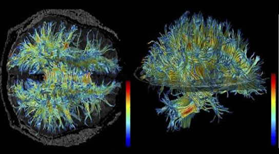
An MRI image of the brain shows the structure of myelin-sheathed wiring (white matter). A new study showed that white matter changed in older subjects to allow for learning, while younger subjects learned via changes in gray matter. Credit: 3D Slicer/Wikimedia Commons
New research from Brown University shows that older people can learn a visual task just as well as younger ones, revealing that a significant change in the white matter of the brain takes place when the older subjects learn.
Providence, Rhode Island (Brown University) — A widely presumed problem of aging is that the brain becomes less flexible — less plastic — and that learning may therefore become more difficult. A new study led by Brown University researchers contradicts that notion with a finding that plasticity did occur in seniors who learned a task well, but it occurred in a different part of the brain than in younger people.
When many older subjects learned a new visual task, the researchers found, they unexpectedly showed a significantly associated change in the white matter of the brain. White matter is the brain’s “wiring,” or axons, sheathed in a material called myelin that can make transmission of signals more efficient. Younger learners, meanwhile, showed plasticity in the cortex, where neuroscientists expected to see it.
“We think that the degree of plasticity in the cortex gets more and more limited with older people,” said Takeo Watanabe, the Fred M. Seed Professor at Brown University and a co-author of the study published in Nature Communications. “However, they keep the ability to learn, visually at least, by changing white matter structure.”
The study’s lead authors are Yuko Yotsumoto of the University of Tokyo and Li-Hung Chang of Brown University and National Yang Ming University in Taiwan. The corresponding author is Yuka Sasaki, associate professor (research) of cognitive, linguistic, and psychological sciences at Brown University.
Spotting the differences
The team’s study enrolled 18 volunteers aged 65 to 80 and 21 volunteers aged 19 to 32 to learn and perform an abstract visual perception task in the lab over the course of about a week. They saw screens showing a background texture of lines oriented in a particular direction. Sometimes a small patch of the screen would quickly show lines pointing in one of two different directions against that background. Subjects simply had to push a button indicating they saw a patch with a particular orientation.
Individuals varied, but older subjects were just as likely on average as younger ones to make substantial progress in discriminating the small patch’s different texture. But the researchers weren’t just interested in whether learning occurred. They also scanned the brains of the volunteers at the beginning and the end of the week using magnetic resonance imaging, which can indicate plasticity in the cortex, and using diffusion tensor imaging, which can indicate changes in white matter.
The scans focused on the section of the brain responsible for visual learning, the early visual cortex (gray matter), and on the white matter beneath it. Moreover, the researchers strategically positioned the texture patch in the same part of the subject’s visual field. That was to ensure that a specific part of the visual cortex (and white matter beneath) that handles signals for that section of the visual field would be trained, while other sections would not.
In analyzing the scan results and the learning performance results together, the researchers found several important associations:
- For changes in the cortex, younger learners showed significantly more than older learners. For changes in white matter, older learners showed significantly more than younger learners.
- In volunteers of both age groups, brain changes occurred only in the sections corresponding with the specific part of the visual field where the patches occurred.
The study produced another curious finding. In looking more deeply at the association between white matter changes and learning performance in the older subjects, the researchers found that they separated into two clearly distinct groups: “good learners” and “poor learners.” In the group that learned very well (their accuracy in discriminating the patch increased by more than 20 percent), members showed a positive association between white matter changes and their improved learning. But among the “poor learner” group (which had a less than 20 percent improvement), the trend was that learning improvement decreased with greater white matter change.
The study doesn’t explain what accounted for why older subjects fell into one group or the other.
The results also don’t definitively explain why white matter plasticity would enable good learners to learn well, although improved signal transmission efficiency is one hypothesis.
But for many seniors, it may be encouraging to learn that plasticity doesn’t necessarily decline with age, it may just shift with the whitening of hair to the brain’s white matter.
Reference: “White matter in the older brain is more plastic than in the younger brain” by Yuko Yotsumoto, Li-Hung Chang, Rui Ni, Russell Pierce, George J. Andersen, Takeo Watanabe and Yuka Sasaki, 19 November 2014, Nature Communications.
DOI: 10.1038/ncomms6504
In addition to Watanabe, Sasaki, Yostumoto, and Chang, other authors are Rui Ni of Wichita State University, and Russell Pierce and George Andersen of the University of California–Riverside.
The National Institutes of Health supported the research.


In Alzheimerism, which is the disease of the old, we know very well there would be only short memory loss and never the long memory loss, say for example language and numerical skilss. This is because the ring of Hippocampus which lies in the mid brain forming a girdle is the immediate seat for all the recent memories. As the files become full they are transferred to parietal region of the brain to be stored in the hard disk. Thus for the oldies there is more transfet of data to parietal region of the brain which is close to optical chiasma or regioin of the brain. Naturally for the older people, the white matter should also come into play and it is not meant only for scaffolding of the brain. For younger people, there is more scope for the grey matter to expand its area and transfer of data also will take place slowly. It is also a sort of adaptation to the age. Thank You.