
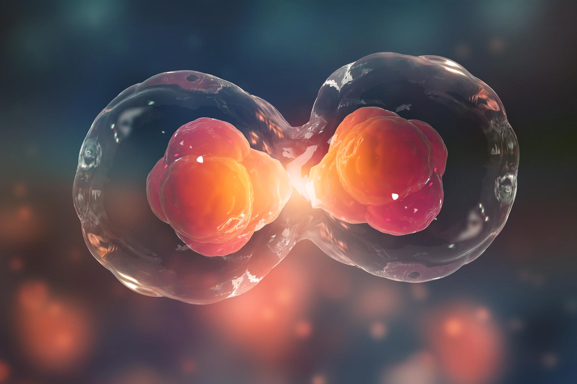
A new study identifies the molecular switch that determines when aging cells permanently stop dividing.
Every time a human cell divides, the tips of its chromosomes get a little shorter. Those tips, called telomeres, act like protective caps. Once they shrink too far, the cell treats the exposed chromosome ends as DNA damage and stops dividing for good.
That shutdown, known as replicative senescence, is one of the body’s built-in defenses against cancer. By forcing damaged or risky cells into permanent arrest, it can stop early cancer clones before they become full tumors. A new study in Molecular Cell now shows that this safety mechanism depends entirely on ATM kinase, a signaling protein that senses DNA breaks and helps protect genomic stability.
The work also helps explain a puzzle that has bothered cell biologists for decades. Cells grown in standard lab air, which contains far more oxygen than most tissues in the body, stop dividing sooner than cells kept at lower oxygen levels. According to the new findings, high oxygen makes ATM unusually reactive, causing cells to become less tolerant of short telomeres.
“Our results have illuminated the mechanism underlying the aging of human cells through replicative senescence,” says Titia de Lange, head of the Laboratory of Cell Biology and Genetics. “These insights are critical for understanding how this tumor suppression pathway prevents cancer.”
Creating the right conditions
Replicative senescence begins when telomeres can no longer recruit enough TRF2, a shelterin protein that helps hide chromosome ends from the DNA damage machinery. Without sufficient TRF2, telomeres start to look like broken DNA, setting off the signal that brings cell division to a halt.
This matters because short telomeres are not only a sign of cellular aging. They are also a warning that a cell may be entering dangerous territory. By stopping such cells from dividing, replicative senescence helps prevent early-stage cancers from expanding.
“Replicative senescence is a remarkably effective tumor suppressor pathway,” de Lange says. “We know this from patients with long telomeres in which this system does not work properly. These patients can get as many as five different cancers before the age of 70, indicating that in people with normal-length telomeres, the telomere tumor suppressor pathway prevents many cancers.”
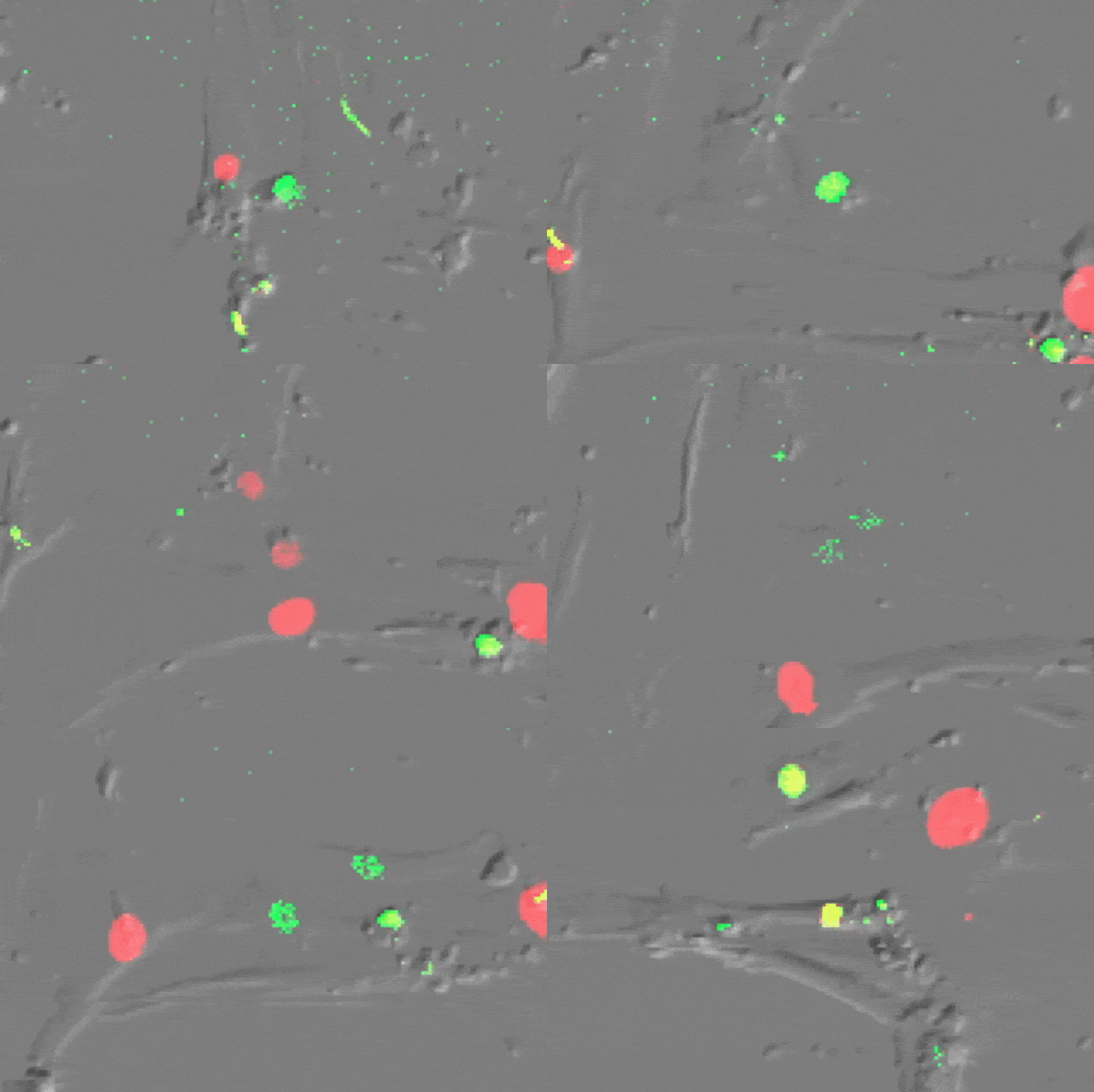
Even so, the exact wiring of this cancer prevention system had remained unclear. Earlier studies suggested that either ATM or ATR, two major DNA damage signaling pathways, might be involved. Oxygen added another complication. Researchers had long known that cells cultured at about 20 percent oxygen reach senescence faster than cells grown at more body-like levels, roughly 1 to 8 percent oxygen. The obvious explanation, that high oxygen simply makes telomeres erode faster, had already been ruled out.
To solve the problem, de Lange and colleagues followed replicative senescence in primary human fibroblasts grown at either 3 percent or 20 percent oxygen. The lower oxygen condition was closer to what many cells experience inside the body, but it also made the experiments much more demanding. Even routine steps such as moving plates, adding reagents, or breaking open cells had to be done quickly to avoid exposing the samples to ordinary air.
“Any time the cells or the reagents are outside of the special low-oxygen incubator, they are exposed to 20 percent oxygen, which can change the molecular environment within minutes,” says Alexander Stuart, a former graduate student in the de Lange lab who now holds a postdoc position at Harvard. “That means you’re often in a race to do all the standard protocol steps extremely quickly so you can keep the samples at low oxygen as much as possible.”
Stuart found that ATM alone was responsible for enforcing senescence at both oxygen levels. When ATM was inhibited, or when TRF2 was overproduced, cells continued dividing past the point where they would normally stop. Blocking ATM signaling in already arrested cells also allowed them to resume growth, showing that the arrest was reversible and fully dependent on ATM.
The impact of high oxygen
The next question was why oxygen changed the timing of senescence so strongly. Stuart and de Lange found that high oxygen does not simply age cells faster in a general way. Instead, it puts ATM into a hyperactive state.
That difference changed how cells responded to extremely short telomeres. At 3 percent oxygen, cells could keep dividing even after many telomeres had become very short. When those same cells were shifted to 20 percent oxygen, ATM responded much more aggressively, treating the short telomeres as serious DNA damage and pushing the cells into senescence.
“I don’t think of it as low oxygen extending the lifespan of human cells—that’s the physiological state of our bodies. Rather, the question was: why do high oxygen conditions shorten cellular lifespan? One could then extend that question: why aren’t high oxygen conditions accurate systems for studying senescence?” Stuart says. “We’ve now shown that high oxygen represents a hyperactive ATM setting, which leads to fewer divisions than cells would naturally undergo.”
The mechanism traced back to reactive oxygen species (ROS), molecules often associated with oxidative stress but found here at higher levels under low oxygen conditions. These molecules caused ATM proteins to link together through chemical bridges known as disulfide bonds. Once locked into these dimers, ATM could no longer respond effectively to DNA breaks or eroded telomeres.
With help from Ekaterina V. Vinogradova, head of Rockefeller’s Laboratory of Chemical Immunology and Proteomics, Stuart and de Lange identified where those disulfide bonds form in ATM. They also showed that one of the bonds is required for oxygen to regulate ATM activity.
Together, the findings show that replicative senescence is controlled by ATM, and that oxygen can strongly alter how that control system behaves. For scientists studying DNA damage responses in cultured human cells, the message is practical: experiments performed at standard lab oxygen may not always reflect the conditions inside the body.
“Studying that in human cells cultured at 20 percent oxygen means you’re basically studying the ATM kinase under hyperactive conditions,” de Lange says. “We’re not saying that everybody should switch to working at low oxygen, because it’s very hard to do, but it may be a good idea to verify that what is observed at 20 percent also holds at 3 percent oxygen.”
The findings may also matter for cancer biology. Many tumors grow in low-oxygen environments that suppress ATM activity. That could allow cancer cells to survive with extremely short telomeres that would normally force growth arrest. Restoring ATM function in such settings may offer a way to push vulnerable malignant cells back into senescence.
“Telomere shortening represents a very important cancer prevention program,” de Lange says. “Questions about how this system works have been at the heart of the work in my lab for years now, and we’ll continue to dig deeper into this pathway.”
Reference: “Attenuation of ATM signaling by ROS delays replicative senescence at physiological oxygen” by Alexander J. Stuart, Kaori K. Takai, Railia R. Gabbasova, Henry Sanford, Ekaterina V. Vinogradova and Titia de Lange, 1 December 2025, Molecular Cell.
DOI: 10.1016/j.molcel.2025.11.006
This work was supported by grants from the NIH to T.d.L. (R35CA210036 and R01AG016642).
Never miss a breakthrough: Join the SciTechDaily newsletter.
Follow us on Google and Google News.
1 Comment
thanks for this