
Recovering whole cells from decades-old preserved tissue could make hospital archives a richer source of molecular data.
Imagine a volume labeled “Encyclopedia of Human Tissue” resting on a table. Inside might be information about how different cells appear in healthy and diseased tissue, how gene activity changes with age, or why cells from different patients respond differently to treatment.
But the book cannot be opened. A plexiglass case seals it shut, leaving its contents visible in principle but inaccessible in practice. Hundreds of similar volumes sit nearby, carrying information that cannot easily be read.
A method developed in the laboratory of Professor of Cell and Developmental Biology Ken Lau could give scientists access to molecular information preserved inside old patient tissue samples that has previously been difficult to retrieve.
Old tissue archives hold untapped data
For more than a century, hospitals and laboratories worldwide have preserved human tissue in formalin (also known as formaldehyde) and embedded it in paraffin wax. This process allows clinicians and researchers to cut tissues into thin sections, stain them, and inspect them under a microscope. But formalin-fixed, paraffin-embedded samples have remained difficult to analyze with some newer sequencing methods.
One example is single-cell RNA sequencing. Known as scRNA-seq, the technology has changed how researchers understand the diversity of cells within tissues by showing that individual cells can produce different types and amounts of RNA—the intermediary between genetic instructions in DNA and the proteins those instructions produce.
The vast archives of FFPE tissue could therefore contain valuable molecular information, but preservation can damage that information and make it difficult to recover, according to Lau lab graduate student James Evans.
Earlier approaches have successfully extracted nuclei from FFPE tissue, but removing the surrounding cell causes substantial information to be lost, especially RNA. In research led by Evans and co-first author Joey Simmons—a senior research specialist in the Lau lab—Lau’s group expanded on a technique Simmons developed to recover more high-quality, intact cells for analysis. Their work was published in Cellular and Molecular Gastroenterology and Hepatology in August 2026.
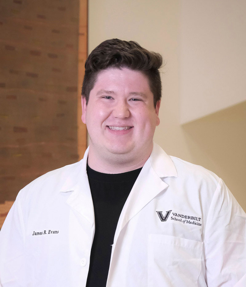
“By recovering whole cells instead of only their nuclei, our method captures more of the biological information contained within each cell. This could make the enormous collections of patient samples already stored in hospitals a source of rich molecular data,” Evans said.
The approach was also particularly effective at retrieving cell types that are often recovered in small numbers, potentially giving researchers a more complete view of the cellular makeup of a tissue.
Benefits vary across different tissues
Evans and his colleagues evaluated the technique using colon and thymus tissue. Whole-cell recovery produced a much greater benefit in colon samples, while improvements in thymus tissue were more limited.
“Our results suggest that recovering whole cells may be especially useful in tissues containing larger cells that have more material outside the nucleus, such as the cells that line the colon, compared to the smaller immune cells in the thymus,” he said.
Archived samples could connect cells to outcomes
Evans hopes the technique will encourage researchers to revisit patient tissues collected over decades. Samples linked to patients whose medical histories and outcomes are already known could be especially valuable.
“Researchers could examine an archived tumor and investigate how the individual cells within that tumor differed between patients who responded differently to treatment or experienced different outcomes,” he said. “Studies like these could eventually help researchers better understand how diseases develop and progress, identify biological features associated with patient outcomes, and discover new markers or potential targets for treatment.”
With the new technique, vast archives of preserved tissue that have long been difficult to study at the level of whole cells could become accessible to modern molecular analysis.
Reference: “Intact Cell Dissociation of FFPE Colon Tissue Enhances Cell Recovery and Gene Detection in scRNA-seq” by James R. Evans, Alan J. Simmons, Zhengyi Chen, Monica E. Brown, Matthew E. Bechard, Morad C. Malek, Cody N. Heiser, Frank Revetta, M. Kay Washington, Laura P. Hale, Robert J. Coffey and Ken S. Lau, 5 August 2026, Cellular and Molecular Gastroenterology and Hepatology.
DOI: 10.1016/j.jcmgh.2026.101863
This research used funds from the National Institutes of Health and the National Science Foundation.
Never miss a breakthrough: Join the SciTechDaily newsletter.
Follow us on Google and Google News.