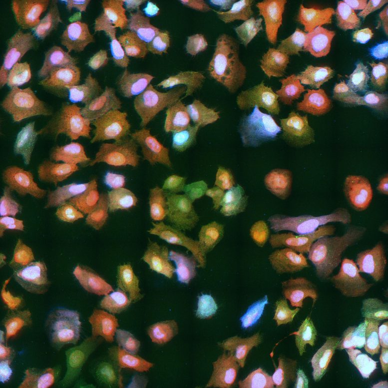
Live-capture of virus infected cells. After infection of a host cell, a virus tries to replicate (green dots), and tries to prevent the host cell from interfering with viral replication by attacking the host cell’s nucleus (blue) and shutting-down protein production of the host cell (red). Credit: Sanne Boersma © Hubrecht Institute
Researchers from the Hubrecht Institute and Utrecht University developed an advanced technique that makes it possible to monitor a virus infection live. The researchers from the groups of Marvin Tanenbaum and Frank van Kuppeveld expect that the technique can be used to study a wide variety of viruses, including SARS-CoV-2 — the virus responsible for the current pandemic. The technique named VIRIM (‘virus infection real-time imaging’) is therefore very valuable for gaining insights in virus infection in the human body. Eventually, this can lead to more targeted treatments for viral infection. The results were published in the leading scientific journal Cell on the 13th of November.
Viruses have a large negative impact on society. This is demonstrated once again by the enormous consequences of the current global outbreak of SARS-CoV-2 for our physical and mental health and for the economy.
Intruder
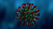
RNA-viruses represent a large group of viruses, which carry their genetic information in the form of RNA: a molecule that is similar to DNA, the genetic material of humans. After infection of a host cell, an RNA-virus hijacks many of the host cell’s functions and turns it into a virus-producing factory. This way, the intruder can quickly replicate inside cells in the body. The new virus particles are subsequently released through the respiratory tract and can infect other people. Examples of RNA-viruses include coronaviruses, the hepatitis C virus, the zika virus, and enteroviruses — a group of viruses that includes rhinoviruses, causing the common cold, coxsackieviruses, that are an important cause of viral meningitis and encephalitis, and the poliovirus, that causes paralytic poliomyelitis.
Upon infection of a host cell, only a single viral RNA molecule is present (spot at start of infection). During replication, the virus replicates the RNA molecule (increase in spots). VIRIM (virus infection real-time imaging) enables the analysis of replication directly from the start of infection. Credit: Sanne Boersma © Hubrecht Institute
Live stream
Until now, available techniques could only provide a snapshot of virus-infected cells. In other words, researchers could see the infected cells at a certain point in time, but it was not possible to monitor the process of virus infection from beginning to end. The newly developed microscope technology VIRIM (‘virus infection real-time imaging’) changes that: researchers from the groups of Marvin Tanenbaum (Hubrecht) and Frank van Kuppeveld (Utrecht University) developed this advanced method with which the entire course of a virus infection can be visualized in the lab with great precision. “This new method enables us to address many important questions about viruses,” says Sanne Boersma, first author of the study.
Fluorescent virus
The method uses SunTag — a technology previously developed by Tanenbaum — in an enterovirus, a group of viruses in which Van Kuppeveld has extensive expertise. The SunTag is introduced into the RNA of the virus and labels viral proteins with a very bright fluorescent tag. Using this fluorescent tag, viral proteins can be seen using a microscope, allowing researchers to see when, where, and how quickly a virus produces its proteins and replicates in its host cell. VIRIM is much more sensitive than other methods: protein production from a single viral RNA can be detected. This allows researchers to track the course of the infection from the very beginning.
Upon infection of a host cell, only a single viral RNA molecule is present (spot at start of infection). In this host cell, the virus fail to replicate, as no increase in spots can be detected. Credit: Sanne Boersma © Hubrecht Institute
Competition
The building blocks of our bodies — cells — have their own defense system to detect and eliminate a virus upon infection. Once a virus enters a cell, a competition arises between the virus and the host cell: the virus aims to hijack the cell to replicate itself, while the host tries very hard to prevent this. Using VIRIM, researchers were able to see the outcome of this competition. They found that in a subset of cells, the host cell won the competition. Boersma: “These host cells were infected by a virus, but the virus failed to replicate.” This triggered the curiosity of Boersma and her colleagues and led to a new experiment.
Achilles’ heel of the virus
The researchers helped host cells by boosting their defense system. As it turned out, the very first viral replication often failed in the cells that had received the boost, which prevented the virus from taking over the host. “The first step in the replication process is the Achilles’ heel of this virus: this moment determines whether the virus can spread further,” Boersma explains. “If the host cell does not manage to eliminate the virus at the very beginning of an infection, the virus will replicate and win the competition.” Boersma and her colleagues used a picorna virus for the development of VIRIM. Members of this virus family can cause diseases ranging from the common cold to severe diseases such as Polio.
Wide variety of viruses
VIRIM enables the identification of the vulnerable phases of a wide variety of viruses. The researchers expect the technique to be valuable for research into many life-threatening viruses, including SARS-CoV-2. Boersma explains: “Understanding viral replication and spreading can help us determine the Achilles’ heel of a virus. This knowledge can contribute to the development of treatments, for example, a treatment that intervenes during a vulnerable moment in the virus’ life. That allows us to create more efficient therapies and hopefully mitigate the impact of viruses on society.”
Reference: “Translation and replication dynamics of single RNA viruses” by Sanne Boersma, Huib H. Rabouw, Lucas J. M. Bruurs, Tonja Pavlovic, Arno L. W. van Vliet, Joep Beumer, Hans Clevers, Frank J. M. van Kuppeveld and Marvin E. Tanenbaum, 13 November 2020, Cell.
DOI: 10.1016/j.cell/2020.10.019


Be the first to comment on "Live Capture: Researchers Develop Virus Live Stream to Study Viral Infections"