
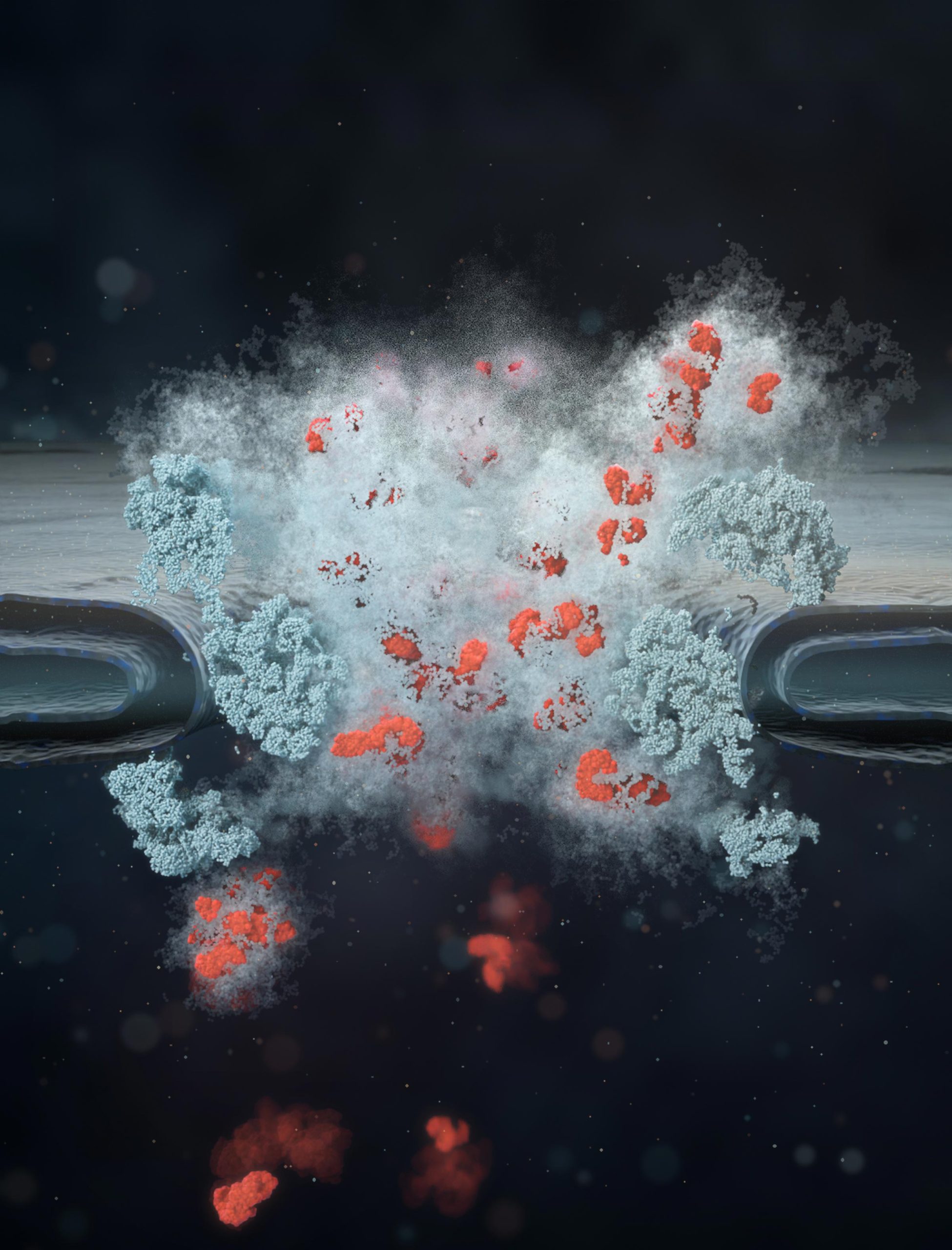
Protecting our DNA takes a surprisingly flexible defense, and scientists have uncovered how cells keep this microscopic barrier fluid rather than letting it harden.
Every minute, tens of thousands of molecules pass through tiny gateways in the membrane surrounding a cell’s nucleus, where its genetic material is stored. Known as nuclear pores, these openings allow certain molecules to enter or leave while blocking others. Scientists have long understood the structure of these gateways, yet the proteins controlling what gets through have remained a mystery. Do they form a rigid barrier, or something more like a constantly shifting liquid?
“As the nuclear pore protects the genome and controls all exchange between the cell nucleus and the cytoplasm, it is one of the most important molecular machines in the human cell,” said Professor Miao Yu of Wuhan University in China, a co-first author of a new study published in Nature. “And also one of the most complex,” added co-first author Maziar Heidari of the Max Planck Institute for Biophysics (MPI-BP) in Frankfurt am Main, Germany.
Living cells keep pore proteins moving
Much of the uncertainty comes from how differently these proteins behave outside living cells. Previous experiments with isolated proteins produced everything from stretched chains to liquid-like droplets and thick gels resembling gummy bears. Some even formed solid deposits.
However, those observations could not establish what happens inside an actual nuclear pore, where thousands of molecular interactions help control traffic. “Such studies did not, however, allow any direct conclusions to be drawn about how the barrier behaves within the living cell,” said Professor Edward Lemke, Professor of Synthetic Biophysics at Johannes Gutenberg University Mainz (JGU).
To investigate the barrier in its natural environment, Lemke and his colleagues attached fluorescent dyes to proteins inside the nuclear pores of living cells. These tiny glowing markers allowed the researchers to track protein movement and determine how flexible the barrier remained. The proteins moved rapidly throughout the entire two-hour observation period, indicating that the interior of the pore behaves more like a fluid than a solid.
“For the first time, we have been able to measure intrinsic protein dynamics within the nanocanal of a living cell and demonstrate that the interior of this canal is fluid-like,” said Lemke, who led the study alongside Professor Gerhard Hummer of MPI-BP. “As the dyes move extremely quickly, the state inside the nuclear pores must be fluid-like,” he explained.
What keeps the proteins from hardening
The contrast became clearer when the researchers compared their observations with proteins isolated in a test tube. After approximately two hours, the protein material consistently hardened into a gummy-bear-like gel or an even more rigid solid. Some of these deposits resembled protein accumulations associated with neurodegenerative diseases such as Alzheimer’s and Parkinson’s. Inside living cells, however, the barrier remained flexible over the same period. “This was a crucial turning point: there had to be something keeping the barrier in the cells dynamic,” said Lemke.
The researchers identified two components that helped explain the difference. One was a transport receptor, a protein involved in moving molecules through nuclear pores. The other was glycosylation, a process that attaches sugar groups to proteins and helps prevent them from sticking together too tightly. When both were included in the laboratory model, the proteins regained much of the mobility observed in living cells. “When we add these two components, we observe a similar mobility in the model system to that found in the nuclear pores of living cells,” said Lemke.
Molecules travel along the pore’s edges
Understanding the barrier’s flexibility also helped the team tackle another longstanding puzzle: where molecules actually travel as they pass through the pore. In an ordinary pipe or river, flow is generally fastest near the center and slower along the edges. Earlier research suggested that nuclear pores work differently, with much of the transport occurring near their outer walls. One proposed explanation was that a biological structure, possibly a plug, occupied the middle and forced molecules toward the sides.
Hummer’s team investigated this possibility using what the researchers describe as the most comprehensive simulation model of a nuclear pore developed to date. The model incorporated experimental measurements from the Mainz team, including how extensively the barrier proteins were linked to one another. It allowed the researchers to examine how molecules would move through a pore with the physical properties they had measured.
“The simulations confirm that the main transport route runs along the edge of the pore,” said Hummer. Importantly, the calculations showed that this pattern could emerge from the pore’s physical and chemical properties without requiring a central obstruction. “While we do not rule out a biological cause such as a plug, it is not necessary from a physicochemical point of view.”
Producing such a detailed model required enormous computing power. “These complex calculations were made possible by JUPITER, Europe’s first exascale supercomputer at the Jülich Research Center,” said Hummer.
The experiments and simulations provide a clearer picture of how nuclear pores maintain a flexible barrier while regulating the movement of molecules into and out of the nucleus. They may also help researchers investigate why proteins that normally remain flexible sometimes harden into deposits, a process involved in neurodegenerative diseases.
Reference: “Glycosylation and importins liquefy the FG barrier in live nuclear pores” by Miao Yu (于淼), Maziar Heidari, Karen Palacio-Rodriguez, Hao Ruan (阮浩), Sara Mingu, Yu Chen (陈宇), Xu-Zhuo Gou (缑绪卓), Pingkai Shen (沈平恺), Songzi Liu (刘松子), Si-Yu Chen (陈思宇), Alam Ahmad Hidayat, Mathis Bode, Mateusz Sikora, Ai-Hui Tang (唐爱辉), Gerhard Hummer and Edward A. Lemke, 11 November 2026, Nature.
DOI: 10.1038/s41586-026-11258-0
Never miss a breakthrough: Join the SciTechDaily newsletter.
Follow us on Google and Google News.