A needle-thin, flexible brain implant is giving researchers a new way to interact with multiple brain regions simultaneously.
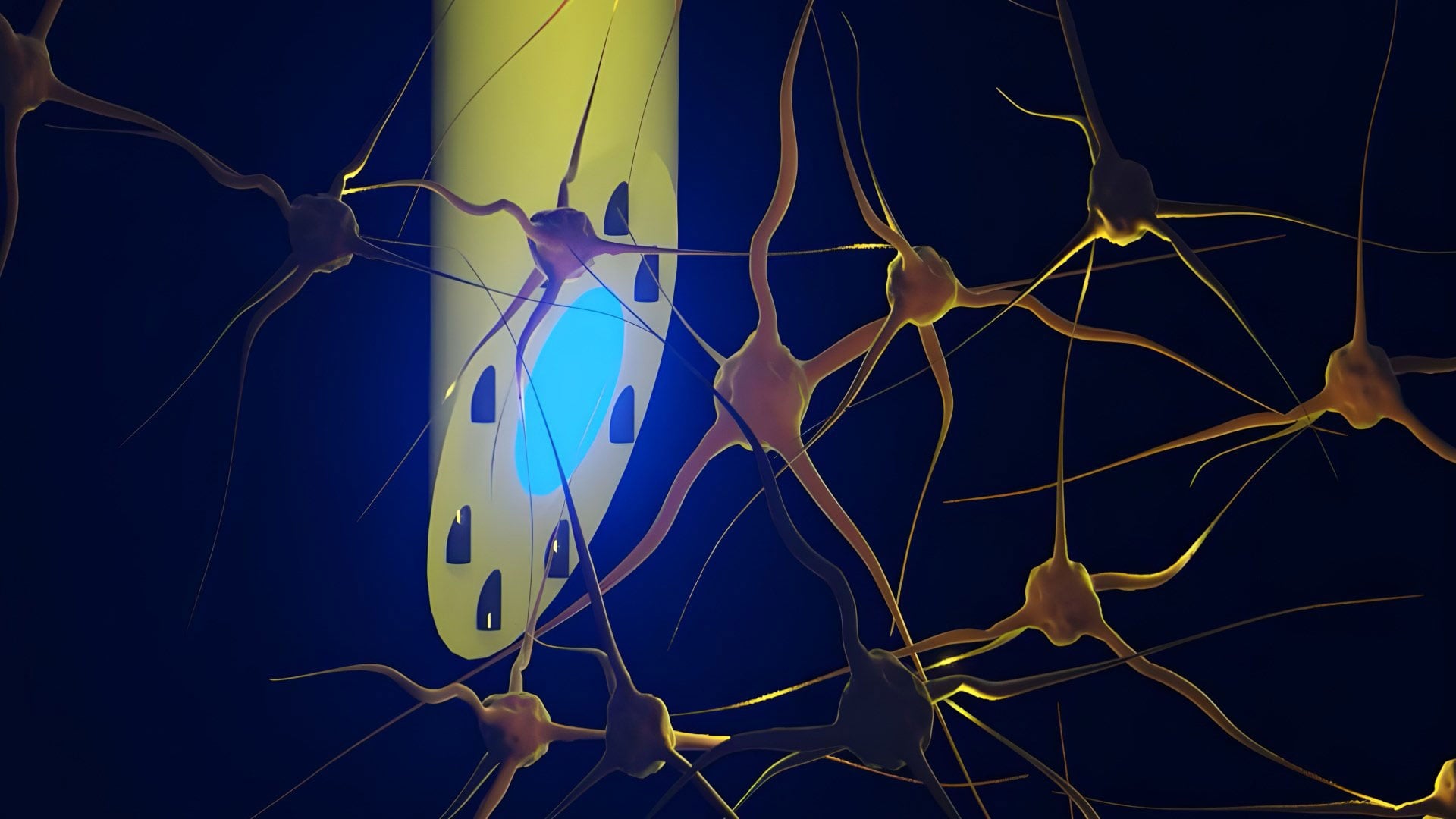
A new type of brain implant is offering researchers a different way to interact with neural circuits across multiple depths of the brain. The device is a microfluidic Axialtrode (mAxialtrode), a long and extremely thin electrode designed to place functional contact points along its length rather than concentrating everything at a single tip. This approach allows scientists to record neural signals while also delivering medication with high spatial precision to different brain regions.
The work was carried out by researchers from DTU, the University of Copenhagen, University College London, and other institutions, and has been published in the journal Advanced Science.
At present, the mAxialtrode is intended mainly for basic neuroscience research. Many brain processes depend on activity that spreads across layers of tissue, making it difficult to study them using tools that interact with only one location at a time. By accessing multiple depths simultaneously, the new implant can help researchers investigate how signals propagate in conditions such as epilepsy and in functions related to memory and decision-making.
The research team also points to longer-term possibilities in medicine. One potential direction involves combining targeted drug delivery with electrical or light-based stimulation in specific brain regions. Using a single implant for both functions could make it easier to study how chemical and physical interventions influence the same neural circuits.
Postdoctoral researcher Kunyang Sui, who developed the mAxialtrode concept together with Associate Professor Christos Markos, explains that the goal was to integrate capabilities that are usually spread across multiple devices. Bringing these functions into one implant can reduce the amount of material placed in the brain while improving experimental control and precision.
A Softer, Less Invasive Implant
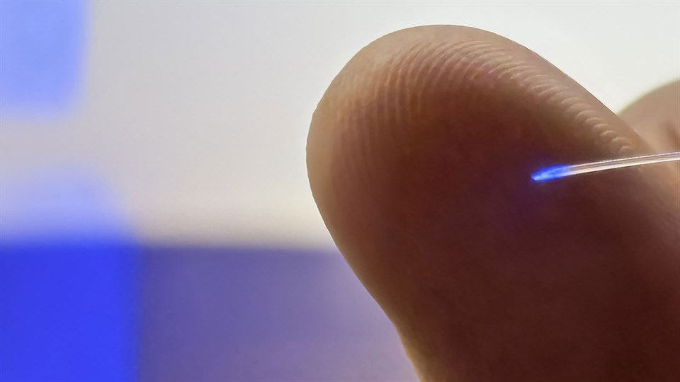
“Most current brain implants are based on hard materials such as silicon, which can irritate the brain and trigger inflammatory reactions in the tissue. The new implant differs in that it is made of soft, plastic-like optical fibers and has a specially angled tip that makes it smaller and reduces the damage caused when it is placed in the brain,” says Kunyang Sui.
He adds that the technology is still at an early stage, with extensive testing, further development, and regulatory approvals required before any clinical use would be possible.
Many neuroscience experiments today rely on conventional flat-end optical fibers. These thin glass or plastic fibers are commonly used to deliver light deep into the brain, including in optogenetics, where light is used to activate nerve cells. Their main drawback is that their influence is limited to a single point.
The outermost end of the fiber is known as the distal tip, or the “nose” of the fiber. Because all light emission and contact with brain tissue occur at this location, researchers can usually stimulate or record activity in only one brain layer at a time, even though many important brain functions depend on interactions across multiple layers and deeper regions.
How the technology is built
The needle-thin mAxialtrode is manufactured using a process in which a large polymer rod is heated and drawn out into a very thin fiber—the process can be compared to making sugar thread, only much more precisely. In the middle runs a core that conducts light. Around it are eight microscopic channels that can carry fluid and also accommodate very thin metal wires for electrical measurements.
The fiber is less than half a millimeter thick and is so flexible that it moves with the brain instead of cutting through the tissue. The difference in stiffness is important because hard implants often trigger inflammatory reactions in the brain over time.
Proven technology
The researchers have not only tested the technology in the laboratory, but also “in vivo”—that is, in mice. Here, the brain electrode was implanted in the brain and connected to light sources, measuring equipment, and small pumps for fluid supply.
The experiments showed that the researchers could stimulate nerve cells with blue and red light, measure electrical activity simultaneously from both superficial and deeper brain layers, such as the cerebral cortex and hippocampus, and inject different substances at different depths, up to almost three millimeters apart. All examinations and stimulations could be performed with a single, lightweight fiber that the animals could carry without any obvious signs of discomfort.
The in vivo experiments and neurophysiological validation were carried out in close collaboration with Associate Professor Rune W. Berg from the University of Copenhagen and Associate Professor Rob C. Wykes from University College London, who contributed expertise in neural circuit analysis and epilepsy-relevant models.
The researchers behind the brain electrode are in the process of patenting the underlying technology and clarifying the possibilities for testing the electrode on patients in a clinical department.
Reference: “Multimodal Layer-Crossing Interrogation of Brain Circuits Enabled by Microfluidic Axialtrodes” by Kunyang Sui, Neela K. Codadu, Daman Rathore, Guanghui Li, Marcello Meneghetti, Anders L. Bøcker, Rune W. Berg, Rob C. Wykes and Christos Markos, 21 January 2026, Advanced Science.
DOI: 10.1002/advs.202519744
Funding: Lundbeck Foundation Multi-BRAIN, Lundbeck Foundation Multi-BRAIN, EIC Pathfinder project Move2Treat
Never miss a breakthrough: Join the SciTechDaily newsletter.
Follow us on Google and Google News.
1 Comment
You djould let me know I have half a brain other halfs a dead cell