
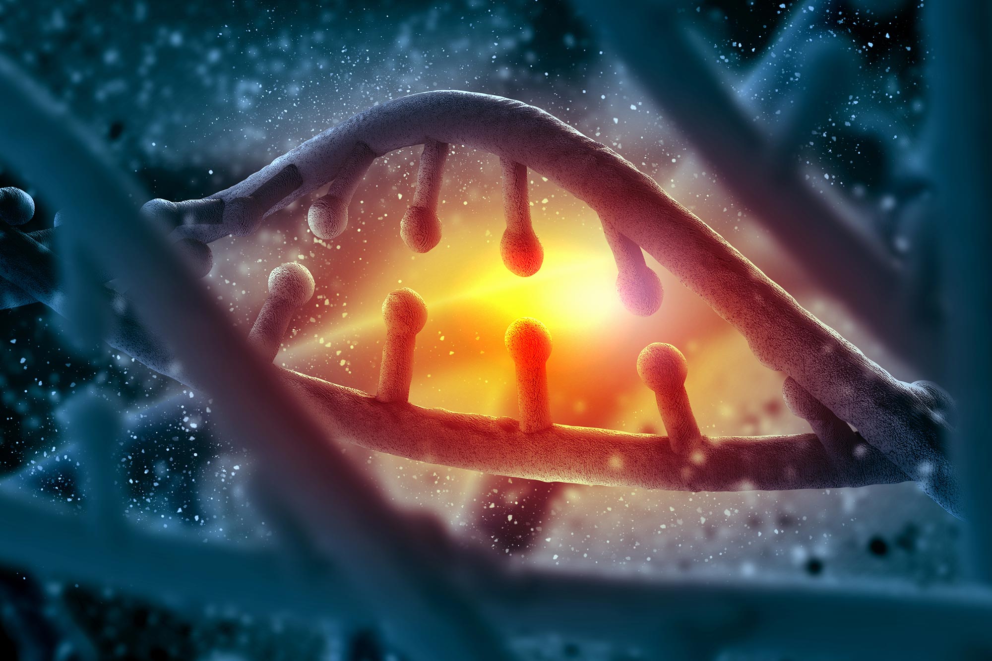
By studying a gene associated with congenital heart defects, scientists uncovered a mechanism showing how small reductions in protein levels can disrupt DNA’s three-dimensional structure and lead to disease.
A baby can inherit one healthy copy of TBX5 and still develop serious heart defects if the other copy does not work. That genetic imbalance is one cause of congenital heart disease, the most common birth defect, which affects about 1 in every 100 babies born each year.
Why a single faulty copy can disrupt heart formation so severely has remained difficult to explain. The working copy continues producing TBX5 protein, yet half the normal amount is apparently not enough.
Researchers at Gladstone Institutes have now uncovered a possible reason. Their study, published in Science, shows that TBX5 helps arrange DNA into the three-dimensional structure required for heart cells to operate correctly. Losing only one copy of the gene was enough to destabilize this organization and alter how numerous other genes were used.
The findings offer a new explanation for haploinsufficiency, a condition in which one functional copy of a gene cannot produce enough protein for normal development.
“TBX5 is just one example of a broader class of genes that cause birth defects when only one copy is lost,” says Benoit Bruneau, PhD, director of the Gladstone Institute of Cardiovascular Disease and a senior author of the study. “What’s exciting about our findings is they suggest many different birth defects might happen for the same reason: the cell’s 3D instruction manual simply gets folded the wrong way.”
“We developed and used different computational models to analyze results from thousands of individual cells,” says Katie Pollard, PhD, director of the Gladstone Institute of Data Science and Biotechnology and the other senior author of the study. “That allowed us to finally see how losing this one protein causes the heart’s DNA structure to break down on every level.”
DNA folding controls cellular instructions
A cell must compress an enormous length of DNA into a space smaller than the head of a pin. It does this by folding the genome into a precise three-dimensional arrangement. Each cell type organizes that structure differently, allowing a heart cell to read a different set of instructions than a brain cell.
This architecture has several levels. Large compartments separate broad regions of the genome, domains organize smaller sections, and chromatin loops bring distant pieces of DNA together. These loops allow regulatory switches called enhancers to physically contact particular genes and activate the instructions a cell needs.

TBX5 guides loops into place
TBX5 was already known as a master regulator of heart development because it activates many genes required for heart cells to form and function. Earlier work from Bruneau’s laboratory showed that losing one TBX5 copy changes the activity of hundreds of heart-specific genes, but the mechanism behind those widespread effects remained unclear.
The researchers asked whether TBX5 controls heart cell function by shaping how DNA folds.
They began with human stem cells carrying either two healthy copies of TBX5, one copy, or no copies. After guiding the cells to develop into heart muscle cells, the researchers used high-resolution 3D mapping to examine chromatin loops and other layers of genome organization.
The experiments generated millions of measurements from thousands of individual cells. Custom computational models allowed the researchers to identify patterns within that immense dataset and compare how different TBX5 levels affected each cell.
“Using the custom computational approaches we developed, we were able to see for the first time how the loss of TBX5 triggers the total collapse of the heart’s 3D DNA organization,” says Shuzhen Kuang, PhD, a first author of the study and former bioinformatics fellow in Pollard’s lab. “Surprisingly, we discovered this collapse happens at every level of genome organization—compartments, domains, and chromatin loops.”
Half the protein disrupts the genome
As healthy stem cells became heart muscle cells, their genomes underwent extensive restructuring. Large DNA regions switched between active and inactive states, creating the organization needed for the developing cells to use heart-related genes.
The results identified TBX5 as a major organizer of this transformation. The protein directs cohesin, a molecular motor that shapes the genome, to particular locations on DNA. Cohesin then creates chromatin loops that connect genes with the enhancers responsible for switching them on.
When TBX5 levels fall, cohesin is not directed to the correct places. Essential loops fail to form, DNA folds incorrectly, and important genes involved in heart development remain inactive.
“What was striking was how the amount of TBX5 matters immensely,” says Zoe Grant, PhD, a first author of the study and a postdoctoral researcher in Bruneau’s lab. “The more TBX5 you removed, the worse the disruption across every level of genome organization we looked at.”

The experiments showed that reducing TBX5 to half its normal level was sufficient to misfold the genome and directly produce heart defects.
The response also varied from cell to cell. Atrial and ventricular heart cells reacted differently to TBX5 loss, and researchers found variation even among individual cells belonging to the same type.
“This could help explain why people with the same mutation can have different heart defects,” Grant says.
The mechanism may extend beyond heart disease
Although the research focused on congenital heart disease, the same mechanism may help explain other developmental disorders caused by losing one copy of an important gene.
“We believe we’ve uncovered a new mechanism of disease,” says Bruneau. “We showed that even a small decrease in a single protein can cause the DNA blueprint to fold incorrectly and lead to disease. So, many birth defects currently attributed to genetic mutations may actually be caused by the 3D misfolding of DNA.”
The researchers next plan to determine when TBX5 begins organizing the genome during early heart development. They will also investigate whether other proteins associated with birth defects control DNA architecture in similar ways.
Reference: “Dose-dependent sensitivity of human three-dimensional chromatin to a heart disease–linked transcription factor” by Zoe L. Grant, Shuzhen Kuang, Shu Zhang, Abraham J. Horrillo, Zhe Chen, Kavitha S. Rao, Cemre Celen, Vasumathi Kameswaran, Carine Joubran, Pik Ki Lau, Keyi Dong, Bing Yang, Weronika M. Bartosik, Nathan R. Zemke, Bing Ren, Deepak Srivastava, Irfan S. Kathiriya, Katherine S. Pollard and Benoit G. Bruneau, 23 July 2026, Science.
DOI: 10.1126/science.adv5434
The work was supported by the National Institutes of Health (NHLBI U01 HL157989, UM1HG011585, R01 HL155906), the California Institute for Regenerative Medicine, Additional Venture, Gladstone Institutes, the Roddenberry Foundation, the Younger Family Fund, UC San Francisco, the Saving tiny Hearts Society, and the National Science Foundation.
Never miss a breakthrough: Join the SciTechDaily newsletter.
Follow us on Google and Google News.