Innovative imaging techniques have allowed scientists to observe and control the synthesis of platinum-nickel nanoparticles, enhancing material design for technology and medicine.
Metallic nanoparticles, made up of a few to several thousand atoms or simple molecules, are gaining considerable attention for their technological potential. Nanoparticle-coated electrodes, known as nanolayers, are especially valuable in fields like energy production, where they function as catalysts. A widely used method for creating these layers on electrodes is electrodeposition. Recently, an international team of researchers uncovered new insights into the complexities of this process.
Nanoparticle Research Advancements
Nanoparticle research is unlocking exciting advancements in energy, medicine, and electronics. A major challenge in this field is controlling how nanostructures are synthesized and grown. To address this, an international team of scientists, led by researchers from the Institute of Nuclear Physics of the Polish Academy of Sciences (IFJ PAN) in Krakow, conducted a groundbreaking experiment.
They demonstrated the electrodeposition of a platinum-nickel (PtNi) nanolayer on an electrode. Using cutting-edge imaging techniques, the team observed the formation of structures at the atomic level in real time—a crucial step toward designing materials with precisely tailored properties.
Understanding Electrodeposition
Electrodeposition is a rapid and convenient method for producing nanostructures. It involves immersing an electrode in a metal salt solution, from which the layer is to be grown, followed by applying an appropriate voltage that causes ions near the electrode surface to reduce, initiating layer growth.
Transmission electron microscopy (TEM) techniques are essential to closely examine the process of electrodeposition. TEM allows for imaging materials with sub-angstrom resolution (i.e., less than one ten-millionth of a millimeter) since it uses an electron beam with a much shorter wavelength than visible light. Ideally, it would be possible to observe, in real-time, how nucleation (the initial growth stage where nanoparticle seeds form) and layer growth occur on the electrode.
However, TEM imaging comes with certain limitations: the samples need to be as thin as possible and entirely dry. To overcome these challenges and enable the imaging of chemical reactions, the researchers utilized thus a special imaging technique in a liquid cell flow chamber.
Advancements in Imaging Techniques
“The flow cell consists of two silicon chips equipped with a 50-nanometer-thick SiNx membrane. This membrane is electron-transparent, and an additional electrode is placed on its surface. By applying a voltage, the microscope user can observe how the layer grows on the electrode. Experiments using such a cell require a special holder for flow experiments in the TEM,” explains Prof. Magdalena Parlińska-Wojtan, Ph.D., Eng. (IFJ PAN).
Real-Time Observations and Insights
Experiments conducted at the Silesian University of Technology using a TEM microscope confirmed that the PtNi layer indeed grows directly on the electrode, providing crucial insights into the fundamentals of the entire process. An alternative mechanism would involve nanoparticles first forming in the electrolyte and then drifting toward the electrode to attach. This effect was also observed, but only in areas illuminated by the beam, due to the fact that the electron beam interacts with water, behaving like a reducing agent.
Subsequent ‘dry’ observations revealed that the layer is actually composed of spherical nanoparticles with diameters of several tens of nanometers. Further magnification of TEM images showed that the surface of these nanoparticles consists of densely branched, fine dendritic structures (multiple branching).
Enhancing Electrodeposition Processes
“As part of our collaboration with the Fritz Haber Institute of the Max Planck Society in Berlin, we conducted an additional experiment by extending the reaction time and reducing the rate of voltage changes. This allowed us to observe additional effects: the nucleation of individual nanoparticles, which rapidly grow and merge to form a continuous layer. During voltage changes in subsequent electrodeposition cycles, the nanoparticles undergo alternating growth and dissolution. However, growth is a faster process than dissolution, which ultimately results in a stable layer,” explains Prof. Parlińska-Wojtan.
Exploring Different Imaging Modalities
As part of the research, another experiment was conducted in liquid environment using a different, but also unique, apparatus: a scanning transmission X-ray microscope (STXM), available at the National Synchrotron Radiation Center SOLARIS in Kraków. During STXM imaging, X-ray radiation is used.
The resulting images do not have as high a resolution as the ones from electron microscopy, but they reveal other properties of the materials under study, such as the oxidation states of atoms in nanoparticles. The result of electrodeposition is not always pure metal; sometimes it is a metal oxide. Depending on whether it is a metal or an oxide (and the oxidation state of the oxide), materials absorb X-ray radiation at different energies.
An STXM image taken with the appropriate energy beam allows for a detailed investigation of the produced nanoparticles. The STXM microscope at the SOLARIS center in Kraków also enabled an experiment in a liquid environment using a flow cell nearly identical to the one used in the TEM. The authors thus performed PtNi electrodeposition inside the STXM and, in real time, investigated the range of X-ray absorption by the nanoparticles. In this way, they determined that the layer actually consists of nickel(II) oxide and metallic platinum.
Significance of the Research
“Conducting an experiment using microscopic techniques in a liquid environment is quite a challenge. Nevertheless, our team succeeded in producing the expected PtNi layer using two different techniques, and the obtained results were complementary,” says Prof. Parlińska-Wojtan.
She emphasizes that “Such research is important for several reasons. The technical reason is that we are still exploring the capabilities and limitations of relatively new, high-end measurement tools. There was also a more important scientific reason: understanding the fundamental factors that govern the synthesis, growth, and properties of nanostructures. This knowledge may help in the future in the fabrication of nanostructured materials tailored better for applications such as fuel cells or medicine.”
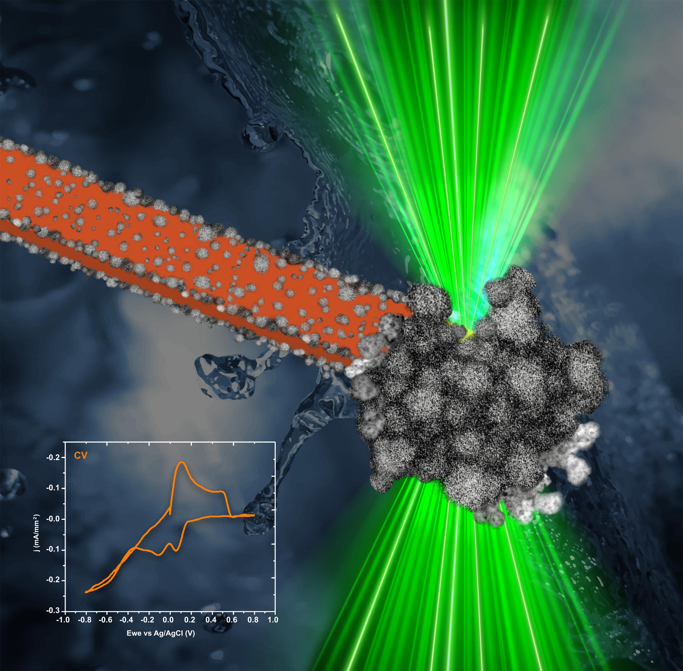
The research results were published in Nano Letters and the journal’s editorial board recognized their work by featuring the accompanying graphic on the cover of one of their issues.
Reference: “Understanding the Growth of Electrodeposited PtNi Nanoparticle Films Using Correlated In Situ Liquid Cell Transmission Electron Microscopy and Synchrotron Radiation” by Magdalena Parlinska-Wojtan, Tomasz Roman Tarnawski, Joanna Depciuch, Maria Letizia De Marco, Kamil Sobczak, Krzysztof Matlak, Mirosława Pawlyta, Robin E. Schaeublin and See Wee Chee, 15 August 2024, Nano Letters.
DOI: 10.1021/acs.nanolett.4c02228
Never miss a breakthrough: Join the SciTechDaily newsletter.
Follow us on Google and Google News.