Silver nanoparticles provide a more efficient way to cut and assemble DNA, improving recovery rates and boosting DNA joining efficiency for future gene engineering applications.
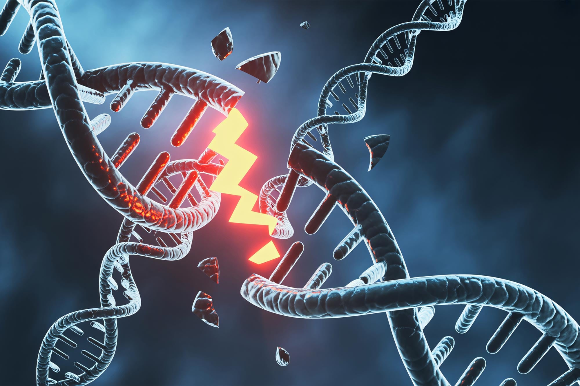
DNA is made of long chains that carry the instructions living things need to grow and function. In genetic engineering, researchers cut DNA at chosen locations and attach the pieces to other DNA sequences. This makes it possible to improve crops, treat genetic diseases, and create animal models used in drug discovery.
To assemble short DNA pieces, scientists often rely on sticky ends, which are short overhanging sequences that help DNA fragments attach more easily. Creating these sticky ends, however, requires highly accurate cutting at specific locations, which remains difficult with existing methods.
A research team in Japan has created a silver nanoparticle-based technique that can cut and reconnect DNA at targeted sites. The method improved DNA assembly efficiency by two to five times compared with standard restriction enzyme approaches. The results were published in Nucleic Acids Research.
Conventional long-chain DNA assembly typically uses restriction enzymes to cut DNA and T4 DNA ligase to reconnect the pieces. The problem is that restriction enzymes recognize only certain sequences and often produce sticky ends that are too short, which reduces joining efficiency.
Replacing Restriction Enzymes with Chemical DNA Cleavage
To overcome this issue, Professor Hiroshi Abe and Assistant Professor Masahito Inagaki of Nagoya University, working with Professor Natsuhisa Oka of Gifu University, investigated whether chemical reactions could cut DNA at desired sites instead of relying on restriction enzymes.
The team focused on a reaction reported between 1990 and 1992, in which silver ions cut 3′-thiol-modified DNA at specific points. They tested whether this reaction could create useful sticky ends. Although silver ions cut DNA effectively, they also attached nonspecifically and caused precipitation. As a result, only about 14% of the DNA was recovered, far too little for practical use.
The researchers then turned to silver nanoparticles. They reasoned that the nanoparticles could be separated after the reaction by centrifugation, which could improve DNA recovery.
Tests showed that DNA-cleaving efficiency reached about 50% at 70°C (158°F) and almost 100% at 95°C (203°F) within two hours. Those temperatures, however, could damage long-chain DNA.
PEG-Coated Silver Nanoparticles Improve DNA Recovery
To solve this problem, the researchers coated the nanoparticles with polyethylene glycol (PEG), a water-soluble polymer that improves stability and dispersion. With PEG, cleaving efficiency rose from 36% to 92% at 37°C (98.6°F) over 31 hours. “In the end, we optimized the conditions to a practical level and, under ambient temperatures, achieved PEG-modified cleaving efficiency above 91% at 50°C (122°F) within just one to two hours,” stated Inagaki, the study’s first author.
The method also removed unwanted DNA pieces that remained attached to the nanoparticle surfaces. This left the desired sticky-ended fragments in solution and raised the final DNA recovery rate from 14% to 98%.
Silver nanoparticles also made it possible to create DNA fragments with 8-base sticky ends, which are difficult to produce using standard restriction enzymes. When the researchers used T4 DNA ligase to join these fragments, the joining efficiency was about twice as high as with traditional methods. With an 18-base overhang, efficiency reached 44%, compared with 8% for a conventional 4-base overhang, a fivefold increase.
Enhanced DNA Assembly Performance and Cell Testing
To test whether the method could work in a practical setting, the researchers assembled a DNA fragment that encodes green fluorescent protein (GFP) and introduced it into human HeLa cells. GFP expression was successfully detected, showing that the DNA had been assembled accurately.
Inagaki commented, “We believe this technology will be useful for synthesizing genomic DNA, with many possible applications in areas such as mRNA library establishment for cancer vaccines and gene therapy, as well as the development of artificial protein drugs and genome crops.”
He also explained the next step: “We have shown that two DNA fragments can be joined. Now, we need to confirm whether multiple fragments can be joined at the same time—a key step for building genome-scale DNA.”
Reference: “Silver nanoparticle-induced site-specific strand cleavage of chemically modified oligonucleotides for long-chain DNA assembly” by Masahito Inagaki, Mikiya Kase, Haruka Hiraoka, Natsuhisa Oka, Fumitaka Hashiya, Naoko Abe, Yasuaki Kimura and Hiroshi Abe, 11 June 2026, Nucleic Acids Research.
DOI: 10.1093/nar/gkag525
This work was supported by the Japan Science and Technology Agency (JST) (JPMJCR18S1, JPMJCR23N1, JP25H00427, JP24H00737, JP22H02219, JP22K21346 International Leading Research), and Japan Agency for Medical Research and Development (AMED) [JP22gm0010008 (LEAP), JP25ak0101289, JP223fa827 (SCADA), JP243fa827032 (SCADA), JP23bm1223009, JP24ek0109697, JP25ama221315, JP25km0405209, JP25ama221230; JP23fk0210133) and Tanaka Kikinzoku Memorial Foundation [Precious Metals Research Grants 2021 Silver Award to M.I.]. Funding to pay the Open Access publication charges for this article was provided by the Japan Science and Technology Agency.
Never miss a breakthrough: Join the SciTechDaily newsletter.
Follow us on Google and Google News.