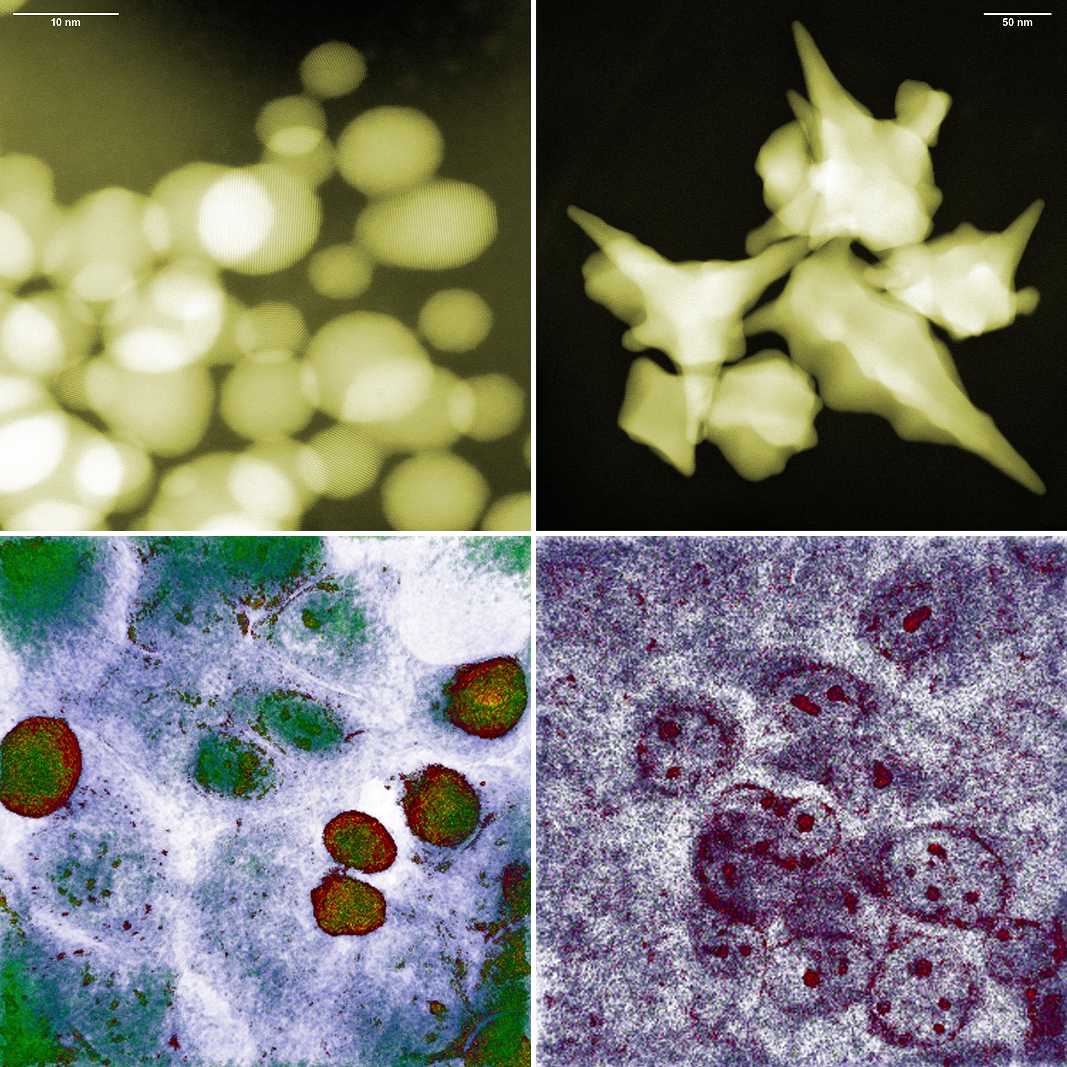
Spherical and star-shaped gold nanoparticles (top) and colon cancer cells after approx. five hours of exposure to them (bottom, respectively). The photo in the bottom left corner proves that, despite the small size of the spherical nanoparticles, the cancer cells survived. False colors. Credit: IFJ PAN
Research overturns old views on gold nanoparticles, showing larger, star-shaped ones are most effective against cancer, leading to a model that improves therapy design.
Scientists have previously assumed that the smaller the nanoparticles used to fight the cancer cells, the faster they die. The logic behind this theory is that small nanoparticles would simply find it easier to penetrate the interior of a cancer cell, where their presence would lead to metabolic disturbances and ultimately cell death.
Now, researchers have used a novel microscopic technique to uncover a more interesting, complex picture of these interactions.
The study, recently published in the journal Nano Micro Small, was conducted by the Institute of Nuclear Physics of the Polish Academy of Sciences (IFJ PAN) and supported by theoretical analysis performed at the University of Rzeszow (UR) and theRzeszow University of Technology.
Colon cancer cells after interaction with small spherical gold nanoparticles did not change their morphology and are still able to divide. Credit: IFJ PAN
Innovations in Nanoparticle Production and Testing
“Our institute operates a state-of-the-art medical and accelerator centre for proton radiotherapy. So when reports emerged a few years ago that gold nanoparticles could be good radiosensitisers and enhance the effectiveness of this sort of therapy, we started to synthesise them ourselves and test their interaction with cancer cells. We quickly found out that the toxicity of nanoparticles was not always as expected,” says Dr. Eng. Joanna Depciuch-Czarny (IFJ PAN), initiator of the research and first author.
Nanoparticles can be produced using a variety of methods, yielding particles of different sizes and shapes. Shortly after starting their own experiments with gold nanoparticles, the IFJ PAN physicists noticed that biology does not follow the popular rule that their toxicity is greater the smaller they are. Spherical nanoparticles of 10 nanometres in size, produced in Cracow, turned out to be practically harmless to the glioma cell line studied. However, high mortality was observed in cells exposed to nanoparticles as large as 200 nanometres, but with a star-shaped structure.
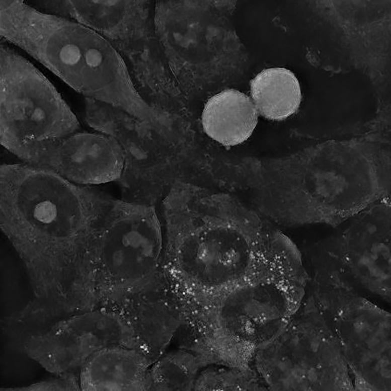
Colon cancer cells after interaction with small spherical gold nanoparticles did not change their morphology and are still able to divide. Credit: IFJ PAN
Holotomographic Microscopy: A Game Changer in Cancer Research
Elucidation of the stated contradiction became possible thanks to the use of the first holotomographic microscope in Poland, purchased by IFJ PAN with funds from the Polish Ministry of Science and Higher Education.
A typical CT scanner scans the human body using X-rays and reconstructs its spatial internal structure section by section. In biology, a similar function has recently been performed by the holotomographic microscope. Here, cells are also swept by a beam of radiation, only not high-energy radiation, but electromagnetic radiation. Its energy is chosen so that the photons do not disturb cell metabolism. The result of the scan is a set of holographic cross-sections containing information about the distribution of refractive index changes. Since light refracts differently on the cytoplasm and differently on the cell membrane or nucleus, it is possible to reconstruct a three-dimensional image of both the cell itself and its interior.
Depciuch-Czarn explained, “Unlike other high-resolution microscopy techniques, holotomography does not require the preparation of samples or the introduction of any foreign substances into the cells. The interactions of gold nanoparticles with cancer cells could therefore be observed directly in the incubator, where the latter were cultured, in an undisturbed environment, what’s more with nanometric resolution, from all sides simultaneously and practically in real time.”
Impact of Nanoparticle Shape on Cancer Treatment
The unique features of holotomography allowed the physicists to determine the causes of the unexpected behavior of cancer cells in the presence of gold nanoparticles. A series of experiments was conducted on three cell lines: two glioma and one colon. Amongst others, it was observed that although the small, spherical nanoparticles easily penetrated the cancer cells, the cells regenerated and even started to divide again, despite the initial stress. In the case of colon cancer cells, the gold nanoparticles were quickly pushed out of them. The situation was different for the large star-shaped nanoparticles. Their sharp tips perforated the cell membranes, most likely resulting in increasing oxidative stress inside the cells. When these cells could no longer cope with repairing the increasing damage, the mechanism of apoptosis, or programmed death, was triggered.
Theoretical Modeling and Practical Applications
“We used the data from the Cracow experiments to build a theoretical model of the process of nanoparticle deposition inside the cells under study. The final result is a differential equation into which suitably processed parameters can be substituted – for the time being only describing the shape and size of nanoparticles – to quickly determine how the uptake of the analyzed particles by cancer cells will proceed over a given period of time,” says Dr. Pawel Jakubczyk, professor at the UR and co-author of the model. “Any scientist can already use our model at the design stage of their own research to instantly narrow down the number of nanoparticle variants requiring experimental verification.”
The ability to easily reduce the number of potential experiments to be carried out means a reduction in the costs associated with the purchase of cell lines and reagents, as well as a marked reduction in research time (it typically takes around two weeks just to culture a commercially available cell line). In addition, the model can be used to design better-targeted therapies than before – ones in which the nanoparticles will be particularly well absorbed by selected cancer cells while maintaining relatively low or even zero toxicity to healthy cells in the patient’s other organs.
Future Directions in Nanoparticle Cancer Research
The Cracow-Rzeszow group of scientists is already preparing to continue their research. New experiments should soon make it possible to extend the model of the interaction of nanoparticles with cancer cells to include further parameters, such as the chemical composition of the particles or further tumor types. Later plans also include supplementing the model with mathematical elements to optimize the efficacy of photo- or proton therapy for indicated combinations of nanoparticles and tumors.
Reference: “Modeling Absorption Dynamics of Differently Shaped Gold Glioblastoma and Colon Cells Based on Refractive Index Distribution in Holotomographic Imaging” by Joanna Depciuch, Paweł Jakubczyk, Dorota Jakubczyk, Bartosz Klebowski, Justyna Miszczyk and Magdalena Parlinska-Wojtan, 15 May 2024, Small.
DOI: 10.1002/smll.202400778


Be the first to comment on "New Research Shatters Old Beliefs About Gold Nanoparticles and Cancer"