
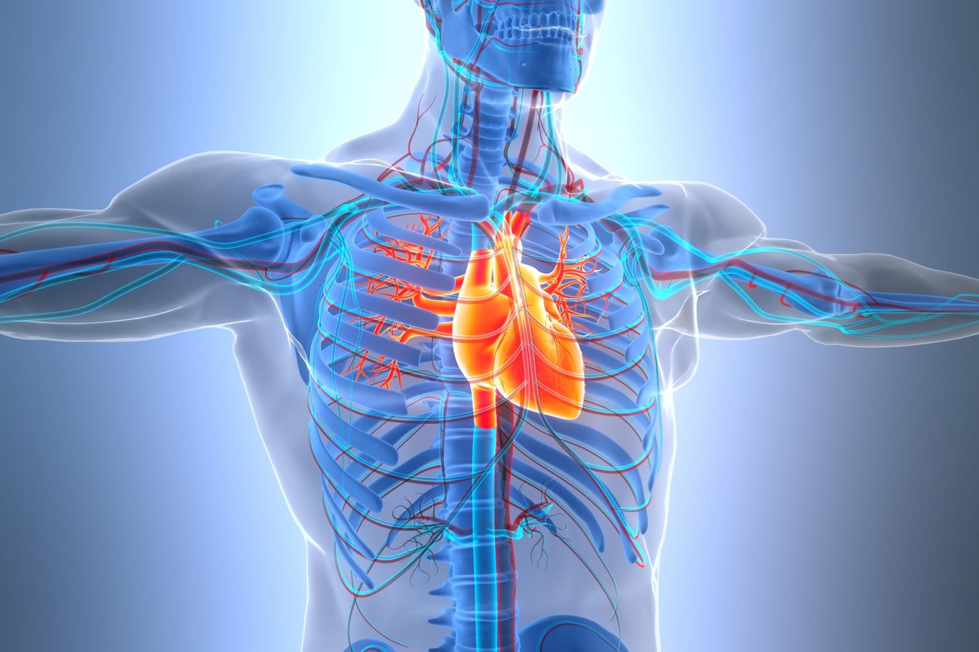
A new study reveals surprising differences in how male and female hearts age at the cellular level.
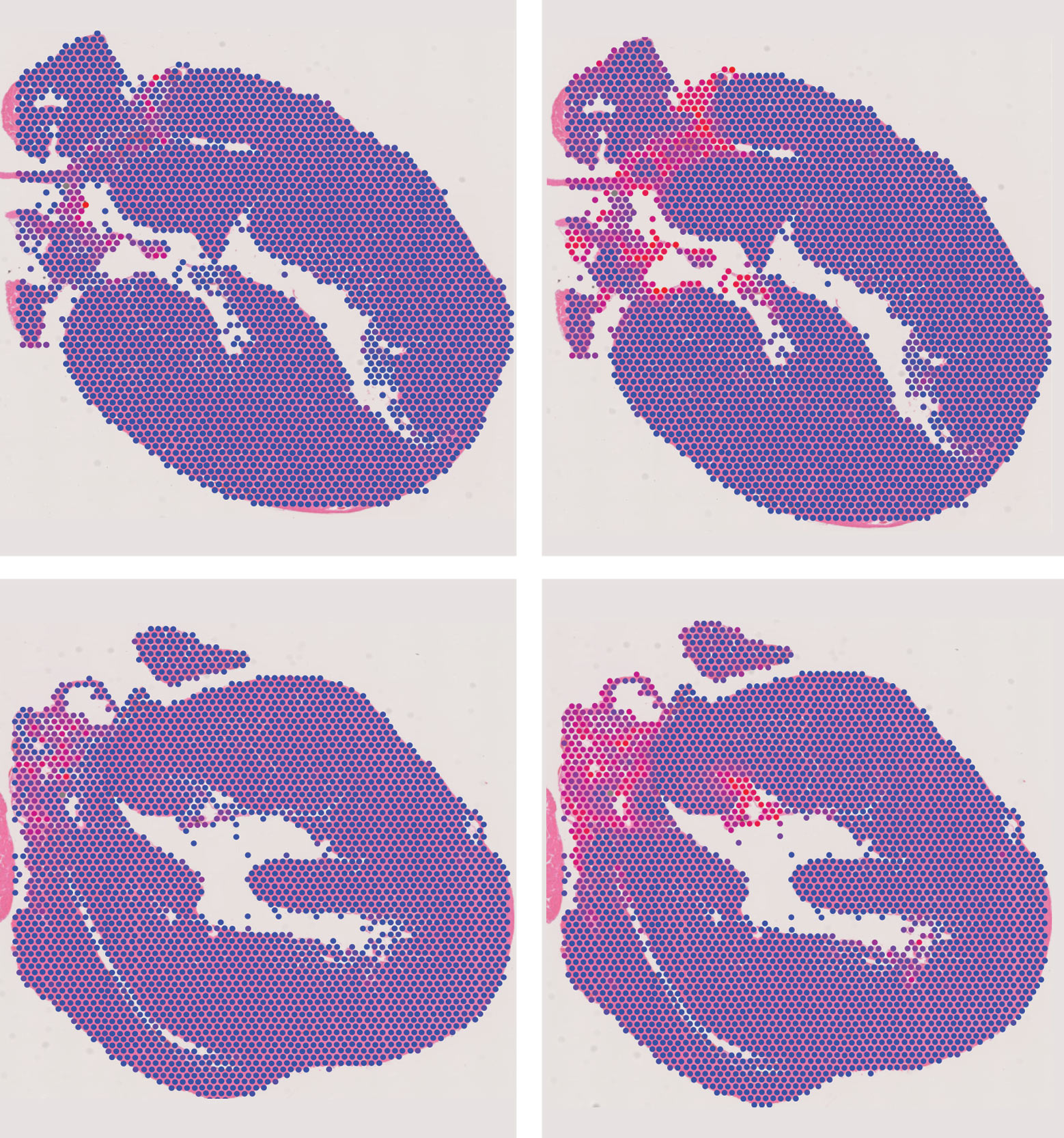
The heart may age very differently from one region to another. A detailed study of mouse hearts found that molecular signs associated with chronic inflammation and tissue scarring were concentrated around the aortic valves and aortic root, rather than being distributed evenly throughout the organ.
Researchers at The Jackson Laboratory (JAX) identified these hotspots by mapping thousands of cells from young and aged mice. Published in Aging Cell, the findings suggest that structures often overlooked in cardiac aging research may play a central role in how the heart changes over time.
Why the Heart’s Valves May Be Aging Hotspots
Previous studies of cardiac aging have largely concentrated on heart muscle, particularly the left ventricle, which pumps oxygenated blood into the aorta and throughout the body. Because mammalian heart muscle cells have very limited capacity for regeneration, understanding how they deteriorate with age has been a major research priority. The JAX team instead examined the entire heart, using single-cell and spatial genomics technologies to identify both the molecular activity of individual cells and their locations within the organ.
“The valves and the major blood vessels are parts often thought of as plumbing rather than the main engine, but this is a very complicated little machine,” said study author Nadia Rosenthal, scientific director at JAX. “It turns out they’re home to many of the cells we’re interested in, and that’s where some of the most important aging-related signals were concentrated.”
“Some of the markers associated with inflammaging were there from the very beginning in young hearts, but they were focused specifically in areas no one had defined before, in the valves and the aorta rather than in muscle tissue,” Rosenthal said. “We really have to think of aging as something that is not linear, because just like we know that people don’t age at the same rate, what we know now is that organs and tissues don’t age at the same rate either, even within one individual.”
How Immune Cells and Fibroblasts Influence Cardiac Aging
Aging is associated with persistent, low-grade inflammation, sometimes called “inflammaging.” The researchers focused on two cell populations involved in this process: immune cells that help regulate inflammation (macrophages) and structural cells responsible for maintaining connective tissue and producing scar tissue (fibroblasts). In older mouse hearts, macrophages showed increased activity in genes associated with inflammation and tissue remodeling, while fibroblasts exhibited molecular patterns linked to fibrosis (or scarring).
“To keep muscle cells happy, the other cells in the heart have to continue supporting them throughout life,” Rosenthal said. “What this study does, for the first time, is provide a comprehensive picture of how those support cells, particularly fibroblasts and macrophages, change with age and contribute to what we call inflammaging.”
The team also detected increased activity in two genes within the identified hotspots. Ccl8 produces an inflammatory signaling molecule, while Cdkn1a (also known as p21) helps regulate the cell cycle and is commonly associated with cellular senescence. Their activity points to changes in signaling between neighboring cells, not simply isolated changes within individual cell types. “We’re not talking about single genes. We’re talking about an entire conversation between immune cells and fibroblasts. As the heart ages, the way those cells communicate changes, and that shift appears to be linked to the inflammation and tissue remodeling we see in older hearts,” Rosenthal said.
A Surprising Discovery in Young Hearts
Some molecular markers traditionally associated with senescent cells were already present in young mouse hearts. Cellular senescence occurs when cells permanently stop dividing, often following stress or damage. Senescent cells can contribute to inflammation and tissue deterioration, but the researchers found that the presence of commonly used senescence markers does not necessarily establish that a cell has entered this state.
“What surprised us was that some of these markers that have been associated with aging were already present in young hearts. They weren’t where anyone expected to find them, and that forced us to rethink what those markers are telling us,” Rosenthal said. “That turns the usual interpretation of cardiac aging on its ear because these markers alone don’t mean a cell is senescent. There’s nothing senescent about a six-month-old mouse heart.”
Male and Female Hearts Show Different Molecular Changes
The study also identified differences in how fibroblasts responded to aging in male and female mice. In males, older fibroblasts showed greater involvement in molecular pathways associated with fibrosis. In females, aging was linked more closely to pathways involved in heart development and protein production. These findings describe differences in gene activity, not demonstrated differences in cardiac function or disease risk.
Rosenthal noted that cardiovascular disease patterns also differ between men and women, with disease risk generally increasing after menopause in women. However, the mouse findings do not establish a mechanism for those human differences. Comparisons with previously published human heart data did identify several shared molecular programs associated with aging, including pathways involved in cellular stress responses.
“Our findings change the focus from aging across the entire heart to specific anatomical hotspots, helping us better understand how inflammation and fibrosis are connected and which regions of the heart are involved in the aging process,” said study author Amalia Sintou, a postdoctoral associate at JAX.
What the Findings Could Mean for Cardiovascular Health
Age is the leading risk factor for cardiovascular disease, but separating changes caused by aging itself from those associated with lifestyle, environmental exposures, and disease remains difficult. Although the study did not establish whether the identified cellular changes directly impair heart function, the controlled conditions in which the mice lived helped researchers examine the biological effects of aging.
“Valve replacement is common in older adults, and one of the big questions is how much of the underlying damage is driven by aging itself,” said study author Aleksandra Aljakna Khan, a postdoctoral associate at JAX. “Because all of the mice in our study lived in the same environment and ate the same diet, we were able to isolate the effects of aging and show that aging alone can initiate some of these changes in the valves and aorta.”
The researchers are now investigating how exercise, diet, and disease influence the interactions between immune cells and fibroblasts. Their goal is to distinguish changes that occur during normal aging from those that may increase the heart’s susceptibility to disease. Although the current findings do not immediately suggest new treatments, the cellular map provides a foundation for studying how inflammation and fibrosis develop in specific regions of the aging heart.
Reference: “Alterations in the Immune-Fibrotic Axis Are Spatially Localized in the Aging Heart” by Aleksandra Aljakna Khan, Amalia Sintou, Yue Zhao, Antonios Somarakis, Harshpreet Chandok, Juliana Alcoforado Diniz, Ramalakshmi Ramasamy, Gretchen T. Clark, Leon Frommer, Sikander Hayat, Brian S. White, Paul Robson, Ron Korstanje, J. Matthew Mahoney and Nadia A. Rosenthal, 5 November 2026, Aging Cell.
DOI: 10.1111/acel.70768
This work was supported by the NIH Common Fund through The Jackson Laboratory Senescence Tissue Mapping Center (JAX-Sen TMC U54 AG079753), with additional support from JAX’s National Cancer Institute-designated Cancer Center (P30 CA034196).
Never miss a breakthrough: Join the SciTechDaily newsletter.
Follow us on Google and Google News.