New single-cell study uncovers diverse immune cells in obese mice.
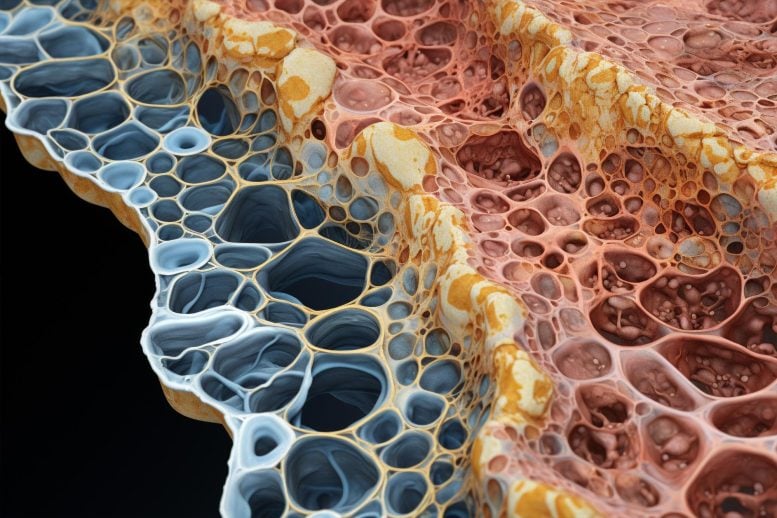
Fat tissue, for as much as it’s been vilified, is an incredibly complex and essential bodily organ involved in energy storage and hormone production, among other functions. Yet, modern lifestyles have led to a worldwide epidemic of obesity, and a corresponding increase in related conditions like type 2 diabetes and cardiovascular disease.
Researchers are attempting to uncover the basics of how fat tissue is structured and, specifically, inflammation associated with obesity, in the hopes of unlocking the connection between the accumulation of fat and poor health outcomes.
A Cutting-Edge Study on Fat Tissue
A new study from Lindsey Muir, Ph.D., Ph.D.-candidate Cooper Stansbury, and their colleagues uses single cell analysis of gene expression combined with spatial transcriptomics to reveal previous unrecognized immune cell types and interactions within adipose tissue. Spatial transcriptomics is a newer technology that captures all gene expression in small spots across an entire thin section of tissue.
Studying fat is easier said than done. In tissues that are organized into defined layers for example the spinal cord or the brain “it’s easier to do sanity checks with your data and identify this or that layer as a particular cell type and know that it should be expressing genes X, Y, and Z,” said Muir, a research assistant professor in the Department of Computational Medicine and Bioinformatics.
“With adipose, it’s a lot more challenging because the cell types are distributed evenly throughout the tissue, without defined cell layers.” In obesity, fat cells, or adipocytes, expand and can reach a limit that ultimately causes cell death and leads to inflammation.
The Experiment: High-Fat Diet in Mice
To better understand the types of immune cells within adipose tissue and where they are in relation to each other in obesity, the team fed mice a high fat diet over the course of 14 weeks, collected fat tissue, and then used single cell and spatial analyses to produce a read out of all the mRNAs present in the sample.
Using a computational process known as clustering in the single cell data, they were able to group cells whose genetic makeup more closely resembled each other than other groups or the overall sample.
They discovered something surprising about the samples’ population of macrophages, an immune cell whose job it is to clean up dead cells and debris.
“We knew going in that macrophages would likely have multiple subtypes… what surprised us were the number that came out that were highly different from each other and coming up at different times and becoming more dominant over time.”
They identified five types, which they named Mac1, 2, 3, 4, and 5. Mac1 was resident to the tissue in both lean mice on a normal diet and obese mice. Mac2 and Mac3, which were identified by their pro-inflammatory genes, peaked after 8 weeks of the high-fat diet.
However, as the high-fat diet progressed to 14 weeks, Mac4 and Mac5 cells, which had low pro-inflammatory gene expression, predominated, while pro-inflammatory Mac2 and Mac3 cells decreased.
“The thinking in the field has been that the type of macrophages that accumulate in obesity are promoting an inflammatory state. Based on these data there’s a lot more to the story,” said Muir.
Her hypothesis is that Mac4 and Mac5 are the lipid associated macrophages (LAMs) described in her own prior work and by other researchers and may be a sign of the body attempting to quell a damaging level of inflammation from pro-inflammatory macrophages and dying adipocytes.
Spatial Transcriptomics: A Closer Look
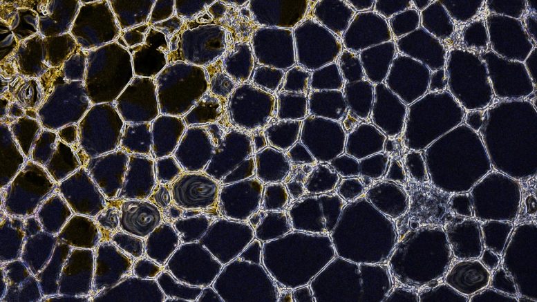
Next, painstakingly careful sectioning of fresh frozen fat tissue enabled analysis by spatial transcriptomics. Each analysis spot in the spatial method has a unique barcode that attaches to the mRNA in the tissue on top of that spot, so gene expression can later be mapped to specific locations in the tissue using the barcodes as coordinates. In this method, the sections are also imaged just prior to collection of mRNA. The study examined these images looking for tell-tale markers called crown-like structures—structures that are associated with insulin resistance.
“Once crown-like structures appear, it takes them a long time to go away and their appearance indicates tissue dysfunction,” noted Muir. “Using image processing, we identified based on the density of these regions what was likely to be a crown-like structure and then went in to verify that we could see them visually,” said Muir. These structures had gene expression indicating the presence of Mac4 and Mac5 LAMs.
With more insight into the cellular makeup and spatial organization of fat tissue in the context of obesity, the next step, Muir said, is to examine the signaling processes and proteins associated with the development of LAMs and metabolic disorders.
Reference: “A lipid-associated macrophage lineage rewires the spatial landscape of adipose tissue in early obesity” by Cooper M. Stansbury, Gabrielle A. Dotson, Harrison Pugh, Alnawaz Rehemtulla, Indika Rajapakse and Lindsey A. Muir, 31 August 2023, JCI Insight.
DOI: 10.1172/jci.insight.171701
Never miss a breakthrough: Join the SciTechDaily newsletter.
Follow us on Google and Google News.