
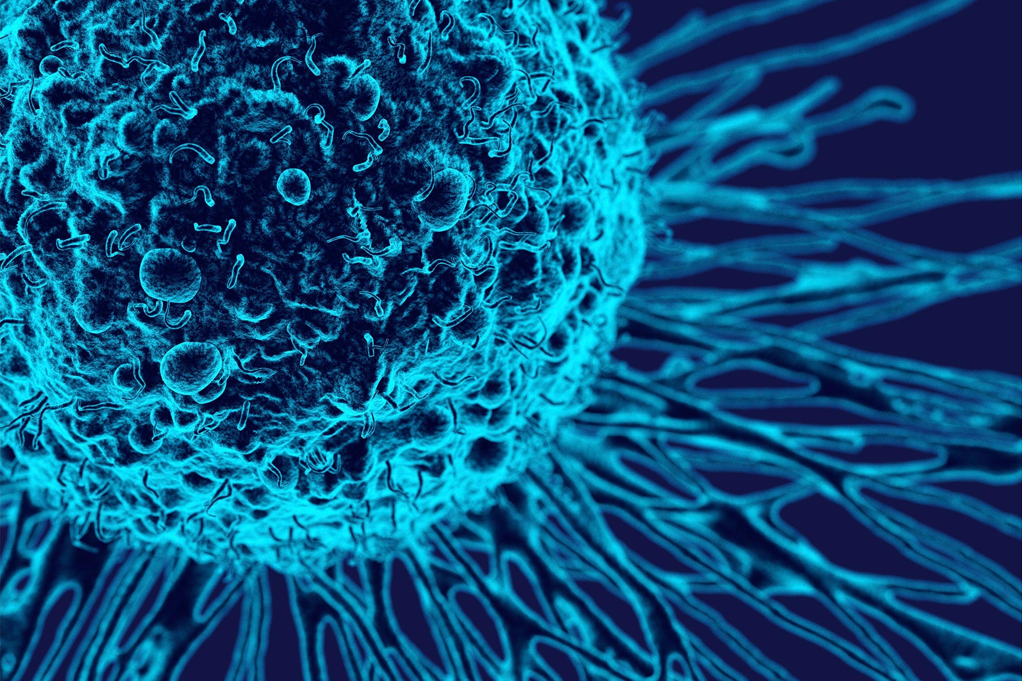
For the first time, new imaging technology in Scotland allows scientists to track cancer from the whole body down to individual cells, providing new insights into the disease.
A medical scan can show where cancer is growing, but not what individual cells inside each tumor are doing. Microscopy can reveal those cellular details, yet it cannot show how the disease is behaving throughout the body. Researchers in Scotland have now connected those two views in a single imaging system.
By combining PET scans, bioluminescence and fluorescence, scientists can locate tumors across the body, identify the most important lesions and then examine those same tumors in detail, including the cells and tissues surrounding them.
Study lead Professor David Lewis of the Cancer Research UK Scotland Institute and the University of Glasgow said: “This exciting technology allows us to build a clearer map of how cancer behaves at both a holistic and microscopic level.
“It allows researchers to follow tumors in the body, identify the lesions that matter, and then zoom in to study those cancer cells and their environment giving us new information about cancer which we can take forward into better and more precise treatments.”
More than 403,000 people in the UK are diagnosed with cancer each year, including 34,800 in Scotland. Around 170,000 people nationwide, including 16,400 in Scotland, die from the disease annually, underscoring the need for better ways to study and treat cancer.
Cancer looks different at every scale
Cancer continually changes. Tumors grow, spread and interact with nearby immune cells and blood vessels. Even within a single patient, separate tumors may respond differently to the same treatment.
Researchers have traditionally examined these processes with separate tools. Whole body methods such as PET scanning can reveal where tumors are located and how they change over time, but they cannot show the behavior of individual cells. Microscopy provides that cellular detail but cannot follow cancer across the entire body.
One system links scans to cells
The Cancer Research UK-funded method brings these two levels of observation together. Connecting large-scale imaging with cellular analysis could help researchers understand why tumors behave differently and why some respond to therapy while others resist it.
Researchers can label cancer cells so they remain identifiable across several imaging methods, allowing scientists to track where the cells travel, how they grow, and how they respond to treatment.
The method also shows how tumor cells interact with their local environment, including nearby immune cells and blood vessels. These surroundings can influence both cancer progression and treatment response.
The technology remains preclinical
The system is currently limited to research in mice and is not available for use in human patients. Even so, it is helping scientists investigate why therapies work against some tumors but not others, test potential treatments more precisely, study individual lesions and develop imaging methods that better represent the underlying biology of cancer.
A Cancer Research UK spokesperson said: “This technology represents a new and powerful tool in our aim to understand cancer biology. Cancer behaves differently from person to person and even tumor to tumor so having the knowledge that allows us to target each with the most effective treatment could be a game changer.
“Being able to see how cancer grows and develops at both a macro and micro level offers us new ways to find more precise ways to tackle and prevent cancer.”
The approach could also strengthen preclinical testing by allowing each tumor to be studied according to its individual biology rather than treating all tumors as identical.
Although researchers demonstrated the method in liver and lung cancer models, they say it could eventually support research across several fields, including oncology, immunology, neuroscience and regenerative medicine.
Reference: “Multiscale in vivo imaging of tumor evolution using a germline conditional triple-reporter mouse” by Ximena L. Raffo-Iraolagoitia, Abdullah Alyamani, Stephanie May, David Stevenson, Agata Mackintosh, Lynn McGarry, Jayanthi Anand, Dmitry Soloviev, Gavin Brown, Colin Nixon, Chrysa Kapeni, Maike De La Roche, Karen Blyth, Thomas Graham Bird, Douglas Strathdee, Scott K. Lyons, Gilbert Fruhwirth, Leo M. Carlin and David Y. Lewis, 15 July 2026, Nature Biotechnology.
DOI: 10.1038/s41587-026-03184-3
Never miss a breakthrough: Join the SciTechDaily newsletter.
Follow us on Google and Google News.