

A softer environment can help struggling tendon cells bounce back, offering a fresh clue to why tendon pain can persist.
Researchers at Queen Mary University of London have identified a population of tendon cells that responds strongly to changes in its physical environment. Their study, published in Advanced Science, offers a new way to investigate tendinopathy, a common condition that can cause lasting pain and restricted movement in people and many animals.
Tendons connect muscles to bones, transmitting the forces that let us walk, run, and lift. But understanding why these tissues deteriorate and why tendon conditions can be difficult to treat requires a closer look at the different cells inside them.
A Softer Layer Between Tendon Bundles
A tendon contains bundles of collagen called fascicles, with softer tissue between them known as the interfascicular matrix (IFM). Cells in these regions occupy different mechanical environments, raising the possibility that they respond differently when those environments change with aging, injury, or disease.
To test that idea, the team isolated and maintained IFM cells alongside cells from the fascicular matrix (FM). Growing both populations on surfaces with different stiffness allowed the researchers to compare their responses under controlled laboratory conditions.
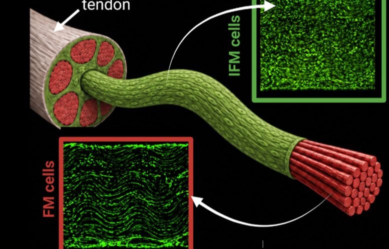
FM cells remained largely unchanged, while IFM cells altered their internal structure and rapidly changed the activity of genes involved in tendon function and production of the extracellular matrix, the material surrounding cells. On stiff surfaces, they also became less able to multiply.
“Our findings show that different tendon cells can respond very differently to changes in their mechanical environment. This is important because tendons can undergo changes in their structure and stiffness during disease, and we need to understand how individual cell populations respond to those changes,” said lead researcher Dr. Simon Grossemy of Queen Mary University of London.
Tendon Cells Recover In Softer Surroundings
Moving IFM cells back onto softer surfaces resembling their usual environment reversed several effects. Their shape and ability to multiply recovered, and some of the changes in gene activity also reversed.
“The recovery we observed when IFM cells were returned to a softer environment was particularly interesting. It highlights how the physical environment is an important factor in maintaining the behavior and characteristics of these cells,” said principal investigator Professor Hazel Screen.
The recovery suggests that some changes in tendon cell behavior can be reversed by altering their surroundings. Studying this response could help scientists understand how the tissue environment shapes cells’ ability to maintain healthy tendons or contribute to disease.
A New Way To Study Tendon Damage
The study does not establish that IFM cells cause tendinopathy or demonstrate a treatment. Its immediate contribution is an experimental system for investigating those questions, including a defined laboratory culture method that helps preserve important IFM cell characteristics. That matters because removing cells from their natural tissue can itself change their behavior.
The findings could also advance research into tendon injury and recovery in animals by helping scientists compare cellular responses across species. Horses are of particular interest because their tendons withstand substantial forces during movement and athletic activity.
Reference: “Unravelling the Distinct Phenotype and Mechanosensitive Properties of Different Tendon Cell Populations” by S. E. Grossemy, D. E. Zamboulis, N. S. Khatib, M. R. Fazal, C. C. Gains, A. Giannopoulos, T. Hopkins, C. Bevan, Y. Aggarwal, M. M. Knight and H. R. C. Screen, 13 September 2026, Advanced Science.
DOI: 10.1002/advs.77391
Never miss a breakthrough: Join the SciTechDaily newsletter.
Follow us on Google and Google News.