
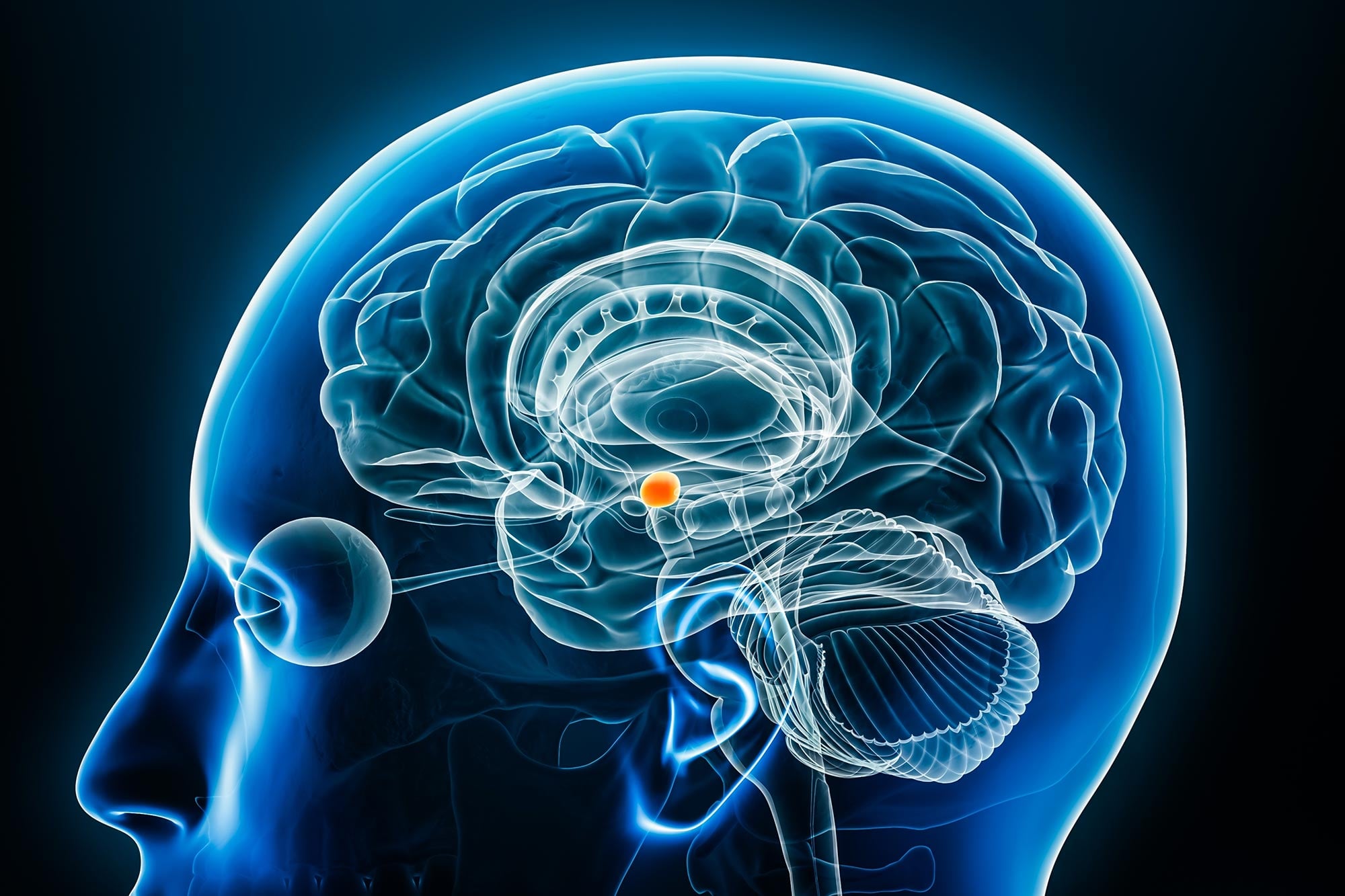
Scientists have discovered that a small brain region best known for processing emotions plays a surprisingly broad role in determining which experiences become lasting memories.
The brain records countless sights and experiences every day, yet only a fraction become lasting memories. Scientists have now identified a pattern of neural activity that helps predict what will be remembered, pointing to a much broader role for a brain region traditionally associated with emotions and fear.
The discovery, made by researchers at Cedars-Sinai Health Sciences University and published in Nature Neuroscience, centers on the amygdala, a walnut-sized structure deep within the temporal lobe. The findings suggest that specialized neurons in this region help determine which new experiences are selected for long-term memory storage.
How the Amygdala Helps Select Lasting Memories
The amygdala has long been known for its involvement in emotional and social behavior, particularly the processing of fear. Its contribution to memory was generally thought to center on emotionally significant experiences. The new research indicates that its influence extends to more ordinary visual experiences, even those without an obvious emotional component.
Researchers identified neurons that respond preferentially to certain types of images, including faces, animals, and landscapes. When these neurons reacted more strongly to an image from their preferred category, participants were more likely to remember that image later. This suggests that the strength of these specialized neural responses helps predict which experiences will remain accessible in memory.
“Our results demonstrate that the activity of specific kinds of neurons in the amygdala predicts, while the person sees an image for the first time, whether that image will later be remembered,” said Ueli Rutishauser, PhD,>
Tracking Individual Neurons as Memories Form
To investigate how the brain selects information for storage, scientists analyzed recordings from 112 patients who had electrodes implanted in their brains to monitor epileptic seizures. These electrodes allowed researchers to measure the electrical activity of individual neurons while participants viewed unfamiliar images.
Later that same day, participants were asked to recall the pictures they had seen. By comparing their memory performance with the earlier neural recordings, investigators could identify patterns of amygdala activity associated with successful recall. The findings were replicated in two separate datasets, strengthening the evidence for the relationship between these neurons and memory formation.
The unusually large collection of single-neuron recordings was assembled through a multi-institutional research consortium led by Cedars-Sinai and supported by the National Institutes of Health (NIH) Brain Research Through Advancing Innovative Neurotechnologies (BRAIN) initiative. Jiayang Xiao, PhD, led the research as the study’s first author.
“This is an impactful study that provides mechanistic insight into the role of the human amygdala—a region commonly associated with emotional responses—in determining which experiences and stimuli become lasting episodic memories. This rigorous human neuroscience study, which uses next-generation tools, has high potential to move discovery toward measurable health impacts,” said Mauricio Rangel-Gomez, PhD, program officer at NIH.
Could the Amygdala Become a Target for Memory Treatments?
The results raise the possibility that disruptions to amygdala activity contribute to memory difficulties associated with neurological conditions. Although the recordings establish a relationship between neuronal responses and later recall, further research is needed to determine whether modifying these responses could improve memory.
“Our findings have important clinical implications, as the memory impairments seen in disorders such as Alzheimer’s disease and temporal lobe epilepsy might be partly due to disrupted processes within the amygdala,” said Rutishauser, also a professor of Neurosurgery. “The amygdala might therefore be a suitable new target for treating these disorders using neuromodulation and/or cellular therapies.”
An additional question is whether the same memory selection process operates across different senses. The current experiments focused on visual information, and Rutishauser said future studies could examine whether the amygdala also helps determine which sounds and smells become lasting memories.
“This study’s insights are the latest in a long line of research led by Cedars-Sinai and collaborators dedicated to revealing how human memory functions,” said Adam Mamelak, MD,>
Reference: “Stimulus-specific recruitment of human amygdala neurons predicts episodic memory encoding success” by Jiayang Xiao, Jonathan Daume, Yousef Salimpour, Natalia Kurilenko, Clayton Mosher, William S. Anderson, Taufik A. Valiante, Adam N. Mamelak and Ueli Rutishauser, 27 October 2026, Nature Neuroscience.
DOI: 10.1038/s41593-026-02493-9
This work was supported by the BRAIN Initiative through the National Institutes of Health (grant no. U01NS117839 to U.R.) and a Center for Neural Science and Medicine Postdoctoral Fellowship by Cedars-Sinai (to J.X.)
Never miss a breakthrough: Join the SciTechDaily newsletter.
Follow us on Google and Google News.