

Scientists are uncovering the secrets of narwhal tusks, revealing an unusual internal structure and new possibilities for studying decades of environmental change in the Arctic.
In the 17th century, King Frederik III of Denmark had a throne made entirely from unicorn horns. The extraordinary royal treasure came from an expedition he sent to Greenland, where the king knew the precious horns could be traced to a very real animal. It lived in Arctic waters, had fins instead of hooves, and was known as the narwhal. The expedition was a success, bringing the Danish ruler a throne that became a symbol of prestige and power across Europe. Yet the legendary horn was not a horn at all. It was a tooth.
And what a peculiar tooth it is. Unlike the curved tusks of elephants and walruses or the incisors of beavers, the male narwhal’s tusk grows forward in an elegant spiral. Usually, it is the left upper canine that extends through the upper lip, twisting counterclockwise as it reaches lengths of up to two meters (6.6 feet), sometimes half the animal’s body length.
That unusual shape has fascinated scientists for years. Now, an international research team has uncovered something remarkable inside the tusk. Its microscopic building blocks form two intertwined spirals that twist in opposite directions, creating a structure that helps the enormous tooth withstand mechanical stress. The findings, published in Nature Communications, offer a new explanation for the anatomy behind one of nature’s most recognizable features.

The strength inside a narwhal tusk
For all their fame, narwhal tusks still hold plenty of mysteries. Scientists do not know exactly how the animals use them, although evidence suggests they play an important role in mate selection and social interaction. Narwhals have occasionally been observed engaging in playful tusk duels, further supporting a possible connection to reproduction and social behavior. The tusks also continue growing throughout the animal’s life and can themselves reach 70 years of age.
Understanding how such a long, spiraling tooth remains strong requires looking far beneath its surface. “Like bones or other teeth in the animal kingdom, the narwhal tusk is a complex composite material whose structure extends from tiny nanoscale building blocks to the visible form of the entire tooth,” says Marianne Liebi, a researcher at the Paul Scherrer Institute (PSI) and co-author of the study.

Most of the tusk consists of dentine, the same material that forms the core of human teeth. A thinner outer layer called cementum covers it, although in most other mammals, this tissue is normally found only around the tooth’s root. Both materials contain collagen fibers reinforced with tiny mineral crystals, combining flexibility with hardness.
The arrangement works somewhat like reinforced concrete. In buildings, concrete withstands compression while a steel framework provides strength against pulling forces. In the narwhal’s tooth, collagen fibers serve as internal reinforcement, while mineral crystals provide hardness. “The narwhal tusk, too, must be both hard and flexible,” Liebi explains. “Only then can it withstand the strong hydrodynamic forces that swimming generates.”
X-rays trace the tusk’s inner structure
The challenge for the researchers was finding out how these microscopic materials relate to the enormous spiral visible from the outside.
“The collagen fibers and mineral crystals are on the nanometer scale, while the spiral of the tooth only becomes visible at centimeter and meter lengths,” Liebi explains.
To connect these vastly different scales, the team turned to a specialized X-ray technique called tensor tomography. Unlike an ordinary dental X-ray, which produces an image based on how strongly tissues absorb radiation, this method uses patterns created when powerful X-rays scatter from regularly arranged structures inside a material.
“From these patterns, we can calculate how the nanometer-sized mineralized collagen fibers inside the material are oriented,” explains first author Adrian Rodriguez-Palomo, who joined the project as a PhD student at Chalmers University of Technology in Sweden and later continued the research as a postdoctoral researcher at Aarhus University in Denmark.
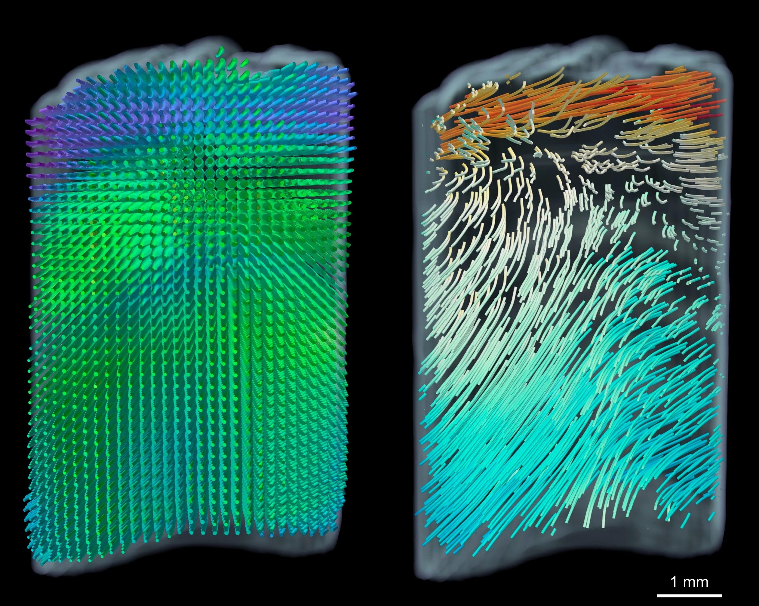
The researchers conducted their investigations at three major European synchrotron facilities: the Swiss Light Source SLS at PSI in Villigen, Switzerland, the MAX IV Laboratory in Lund, Sweden, and the European Synchrotron Radiation Facility (ESRF) in Grenoble, France. These facilities produce the powerful X-rays needed to examine extremely small structures.
During tensor tomography, the tooth is rotated within the X-ray beam and scanned point by point. Millions of measurements are then combined to reconstruct a three-dimensional picture of its internal architecture, connecting the orientation of nanoscale fibers with the structure of the entire tooth.
Two spirals hidden inside one tusk
After collecting this enormous amount of data, however, the scientists encountered an unexpected problem. The spiral they were searching for appeared to be missing.
“That puzzled us quite a bit,” Liebi recalls. Earlier mechanical tests had suggested that the tusk’s distinctive shape should already be reflected in the arrangement of its collagen fibers and mineral crystals. “But instead of a helix, all we saw was a regular pattern.”
The answer emerged when the researchers changed how they examined the data. Rather than concentrating on individual points within the three-dimensional images, they began comparing how those points were oriented relative to one another.
“When we compared how these points were oriented relative to each other, a directional trend began to appear,” recalls Rodriguez-Palomo. “All we had to do then was connect the dots – as in a children’s paint-by-numbers kit – and suddenly the structure became visible: two intertwined spirals.”
The outer cementum layer followed a left-handed helix, while the mineralized collagen structures within the dentine twisted in the opposite direction. Together, these interlocking spirals create an unusually stable architecture, much like the twisted strands of a rope. Their opposing orientations help the tooth resist twisting forces and other mechanical stresses while contributing to its distinctive external spiral.
A lifetime of Arctic climate clues
The researchers are also interested in another remarkable property of narwhal tusks: their ability to preserve a record of the Arctic environment.
Led by Henrik Birkedal of Aarhus University in Denmark, the international team is investigating how tusk growth relates to environmental and climate conditions. Much like the annual rings of a tree, the growth structures within a narwhal’s tooth can provide clues about past conditions, including changes in climate and food availability.
Because the tusk grows continuously, it can preserve environmental information across much of an animal’s life, which can approach a century. A tooth once mistaken for evidence of a mythical creature may therefore also hold a decades-long record of life in the Arctic.
Reference: “The narwhal tusk assembles its macroscopic helix from building blocks with opposing twists” by Adrian Rodriguez-Palomo, Peter Alling Strange Vibe, Dimitra Athanasiadou, Jonas Palle, Leonard C. Nielsen, Jonathan T. Avaro, Thorbjørn Erik Køppen Christensen, Nina Kølln Wittig, Mads Ry Vogel Jørgensen, Innokenty Kantor, Manfred Burghammer, Jiliang Liu, Stefano Checchia, Christian Appel, Karl Anker Jørgensen, Eva Garde, Mads Peter Heide-Jørgensen, Marianne Liebi and Henrik Birkedal, 18 August 2026, Nature Communications.
DOI: 10.1038/s41467-026-75689-z
The study is part of the NordForsk-funded research project “Narwhal tusks – a tale with a twist.” It was supported by the European Union’s Horizon 2020 research and innovation program under the Marie Skłodowska-Curie program.
Never miss a breakthrough: Join the SciTechDaily newsletter.
Follow us on Google and Google News.