
Researchers from Charité – Universitätsmedizin Berlin and the Max Delbrück Center have identified the precise mechanism by which the inflammatory signaling molecule IL-12 contributes to the development of Alzheimer’s disease.
Microglia, the brain’s immune cells, usually act as vigilant protectors. They remove harmful invaders like microbes and clear away cellular debris, including the plaques commonly found in Alzheimer’s disease. However, as the brain ages, microglia begin to change. While some maintain their protective functions, others lose this ability and start releasing low levels of inflammatory molecules.
One of these molecules is interleukin-12 (IL-12). Through detailed analysis, research teams led by Professor Frank Heppner, Director of the Department of Neuropathology at Charité Universitätsmedizin Berlin, and Professor Nikolaus Rajewsky, Director of the Berlin Institute for Medical Systems Biology at the Max Delbrück Center, have uncovered how IL-12 may initiate and speed up the development of Alzheimer’s dementia. Their findings, published in Nature Aging, offer new possibilities for combination treatments.
“For decades, Alzheimer’s research focused almost entirely on amyloid-beta and tau protein deposits, while inflammation was seen as a secondary effect,” says Heppner. “Only recently have we begun to understand that inflammation might be a key factor driving the disease.”
In 2012, Heppner’s lab reported in Nature Medicine that blocking IL-12 and IL-23 significantly reduced Alzheimer ’s-related brain changes in mice. “But we couldn’t determine the exact mechanism using standard methods,” he explains. He suspected that single-cell analysis could provide the answers, so he invited Rajewsky to join the effort.
Sticky and tangled brain cells
Throughout life, cells refer to their genetic instructions to respond to external stimuli. Researchers use single-cell analyses to observe this process, reconstructing which genes are being read and translated into proteins in thousands of individual cells simultaneously.
These analyses generate massive datasets, which can now be analyzed with the help of artificial intelligence and machine learning. However, a major challenge in using single-cell sequencing technology is isolating individual cells from a tissue sample without damaging them or causing unintended changes.
“In aging mouse brains – especially those with Alzheimer’s plaques – cells are so stuck together and tangled that separating them cleanly is nearly impossible,” Rajewsky explains.
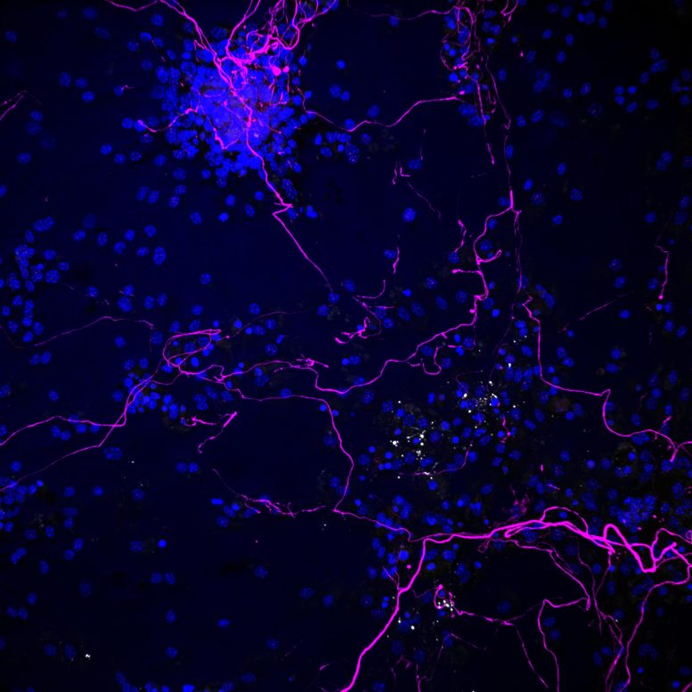
His team spent several years perfecting a workaround. Instead of isolating entire cells, they extract cell nuclei from brain tissue and analyze the RNA present in each cell. By cross-referencing with publicly available data, such as the Allen Brain Atlas, they can ensure that their method provides a representative snapshot of all cell populations. For the present study, they sequenced RNA from over 80,000 cell nuclei and developed specialized workflows to process the data. They also reconstructed communication between cells.
“Our teams repeatedly sat together to try to interpret this highly complex data,” Rajewsky says. “This painstaking early optimization was crucial – without it, we would not have been able to detect these connections.”
How IL-12 damages the Alzheimer’s brain
IL-12, previously known primarily for its role in autoimmune diseases like Crohn’s disease and rheumatoid arthritis, appears to play a pivotal role in Alzheimer’s progression. It damages two key brain cell types: mature oligodendrocytes, which normally produce myelin – the fatty insulating layer around nerve fibers essential for rapid signal transmission; and interneurons, which are particularly important for cognition and memory.
IL-12 binding to interneurons causes them to die. A vicious circle begins: As more microglia produce IL-12, more brain cells sustain damage. Meanwhile, the remaining functional microglia become overburdened by the task of clearing the additional cellular debris, and thus fail to remove Alzheimer’s plaques.
To verify this mechanism, researchers tested it in mice and in human tissue. When Heppner’s team blocked IL-12 in cell cultures and mouse models, they could stem disease-related changes. Electron micrographs of mouse brain tissue taken at the Max Planck Institute for Multidisciplinary Sciences in Göttingen also showed how myelin structure and nerve fiber density changed depending on whether the IL-12 signaling pathway was present or absent.
Mass spectrometric analyses (lipidomics) at the University of Zurich confirmed the altered composition of the fat-rich insulating layer. Study of autopsy tissue from Alzheimer’s patients provided further confirmation of the results – the more advanced the disease, the more IL-12 was present in the tissue. Cell cultures with human oligodendrocytes were also extremely sensitive to IL-12.
Potential combination therapy
“We now have a highly detailed picture of this mechanism, with single-cell technologies serving as a crucial catalyst. The only remaining question is which cell type IL-12 impacts first – oligodendrocytes, interneurons, or both simultaneously,” says Heppner, who is also Group Leader in Neuroimmunology at the Deutschen Zentrums für Neurodegenerative Erkrankungen (DZNE).
The study has immediate implications as there are already drugs on the market that block IL-12. The researchers hope that clinicians will build on their findings and initiate clinical trials. “If these drugs prove effective, they would be a new arrow in the quiver. Alzheimer’s doesn’t just have one cause. One axis of the disease is also controlled by the immune system, at least in some patients. Slowing neurodegeneration will require combination therapy,” Heppner emphasizes. Such an approach could start early in the disease process, as IL-12 can be measured in blood or cerebrospinal fluid, he adds.
Meanwhile, the teams at Charité and the Max Delbrück Center are exploring a new hypothesis: Could microplastic in the brain drive microglia to produce IL-12? “Microglia may struggle to process microplastic, triggering inflammatory reactions,” Rajewsky suggests. “This could reveal a link between environmental factors and widespread diseases.”
While unproven, both teams consider it a compelling and important research direction.
Reference: “Interleukin-12 signaling drives Alzheimer’s disease pathology through disrupting neuronal and oligodendrocyte homeostasis” by Shirin Schneeberger, Seung Joon Kim, Maria N. Geesdorf, Ekaterina Friebel, Pascale Eede, Marina Jendrach, Anastasiya Boltengagen, Caroline Braeuning, Torben Ruhwedel, Andreas J. Hülsmeier, Niclas Gimber, Marlene Foerster, Juliane Obst, Myrto Andreadou, Sarah Mundt, Jan Schmoranzer, Stefan Prokop, Wiebke Kessler, Tanja Kuhlmann, Wiebke Möbius, Klaus-Armin Nave, Thorsten Hornemann, Burkhard Becher, Julia M. Edgar, Nikos Karaiskos, Christine Kocks, Nikolaus Rajewsky and Frank L. Heppner, 13 March 2025, Nature Aging.
DOI: 10.1038/s43587-025-00816-2
Never miss a breakthrough: Join the SciTechDaily newsletter.
Follow us on Google and Google News.