
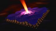
Magnetic order of iron tellurides, imaged with a low-temperature scanning tunneling microscope. The enlarged section shows the atomic structure. Credit: Peter Wahl, University of St Andrews & MPI for Solid State Research
Scientists from the Max Planck Institute for Solid State Research use atomic scale imaging of magnetic structures to study new aspects of high-temperature superconductivity.
Superconductors raise many hopes, especially materials which lose their electrical resistance at quite high temperatures – be it for high-performance medical imaging technologies, the energy transportation, or for maglev trains. High-temperature superconductors which deserve the name could find many applications. However, their fascination bears no relationship to the ongoing mystery of their actual nature; this has so far hindered the search for zero-resistance conductors for realistic temperatures. Scientists from the Max Planck Institute for Solid State Research in Stuttgart and Augsburg are making their contribution to a more detailed understanding of how iron-based superconductors work and the role played by magnetism. They are the first to have imaged the magnetic structure of a so-called strongly correlated electron system, here of iron telluride, on an atomic scale. Prior to this, information about the magnetic structure was provided only by neutron diffraction, but the image it produced was imprecise. Iron telluride is the non-superconducting parent compound of the iron chalcogenide superconductors. The researchers now hope to be able to apply the method to materials which exhibit both superconducting and magnetic properties in order to find out more about the relationship between magnetism and superconductivity.
Substances such as copper oxide ceramics or iron arsenic compounds are deemed to be high-temperature superconductors: they do not have to be cooled as much as other materials in order to render them superconducting. Why is this? To date there are hypotheses, but no proven description of the precise processes. “A key question which many research groups are now posing is the one about the relationship between magnetic and superconducting properties of these materials,” says Peter Wahl from the Max Planck Institute for Solid State Research and University of St Andrews. “Can both effects occur at one and the same location? Or are they mutually exclusive?” Physicists think it is possible that the magnetic properties of the materials could even be the cause of their superconductivity.
In order to examine this, researchers have long been looking for a procedure that allows characterization of the magnetic structures in these strongly correlated electronic materials on the atomic scale. The method of neutron diffraction has so far been the tool of choice for investigating the magnetic order, but it provides only spatially averaged insights into the magnetic structure.
The Stuttgart-based Max Planck researchers have now made use of a so-called spin-polarized scanning tunneling microscope, which can image the orientation of the magnetic moments of individual atoms. The method is not new, but has so far mostly been applied to metal surfaces and nanostructures. What was not clear until now was whether the method could be used to clarify the magnetic structure of a strongly correlated system such as iron telluride. This is because the top layer of this material consists of tellurium, an element which itself is not magnetic.
The scientists have now shown that the spin-polarized scanning tunneling microscope can also be applied to strongly correlated electron materials despite their complex chemistry. The iron lattice below most likely exerts too great an influence. Narrow longitudinal stripes, which result from the anti-ferromagnetic order in the iron telluride, can be recognized in the image taken by the scanning tunneling microscope. Within the stripes, all magnetic moments have the same orientation; on the adjacent stripes, it is in the opposite direction.
An experimental challenge was to magnetize the tip of the microscope for the spin-polarized investigations. To study nanostructures on surfaces, researchers primarily achieved this by heating the tip of the microscope and vapor depositing magnetic material on it. To avoid the need for this technologically demanding procedure, the scientists used a trick: they picked up individual iron atoms on the surface of the iron telluride under investigation with the tip of the microscope, until it became magnetic. In this way, they could image the magnetic stripe order in iron telluride with atomic resolution in real space.
The researchers made an interesting observation on the temperature which is necessary for the anti-ferromagnetic structure to form. In the experiment, this was approximately minus 227 degrees Celsius, around 20 degrees lower than the temperature that is normally necessary. The reason for this is that the researchers observed only the surface of the iron telluride in their experiment. Compared to the iron telluride layers in the bulk of the material, the interactions with an atomic layer above it are missing here. Consequently, the magnetic moments cannot mutually stabilize their order as well – the magnetic structure forms only at a lower temperature.
The Research Group lead by Peter Wahl also determined that the magnetic order becomes more complex when the proportion of iron atoms is higher: the longitudinal stripes partially dissolve, and are overlaid by transverse stripes. It seems that the surplus atoms and their magnetic moments mess up the magnetic. “There is still a great potential for research here,” says Peter Wahl. “I believe that a real boom is going to develop very soon, groups will be carrying out similar experiments on other materials at the boundary between superconductivity and magnetism.” Understanding the properties of these materials would be the first step towards superconducting technology which is more energy efficient, and eventually may even be suitable for routine application.
Reference: “Real-space imaging of the atomic-scale magnetic structure of Fe1+yTe” by Mostafa Enayat, Zhixiang Sun, Udai Raj Singh, Ramakrishna Aluru, Stefan Schmaus, Alexander Yaresko, Yong Liu, Chengtian Lin, Vladimir Tsurkan, Alois Loidl, Joachim Deisenhofer and Peter Wahl, 31 July 2014, Science.
DOI: 10.1126/science.1251682







Be the first to comment on "Atomic Scale Imaging of Magnetic Structures"