Scientists have uncovered new evidence that the gut may play a far more active role in multiple sclerosis than previously recognized.
For decades, multiple sclerosis (MS) has been viewed primarily as a disease of the brain and spinal cord. But growing evidence suggests its origins may begin much farther away: in the gut.
Scientists have increasingly linked changes in the gut microbiome to MS, yet exactly how intestinal microbes influence immune cells that attack the central nervous system has remained one of the field’s biggest unanswered questions.
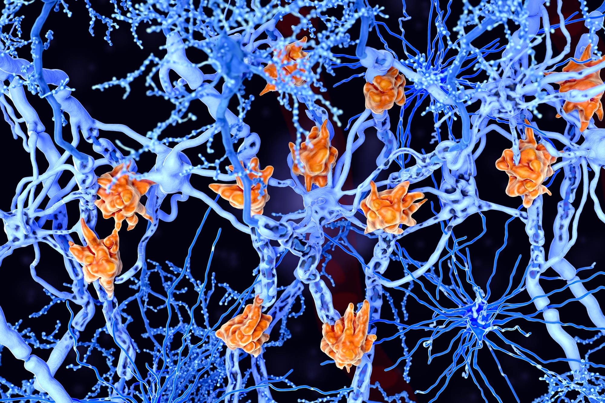
MS is a debilitating autoimmune disease in which the immune system mistakenly attacks myelin, the protective sheath surrounding nerve fibers in the brain and spinal cord. This damage disrupts communication between neurons, leading to symptoms ranging from numbness and muscle weakness to vision problems and cognitive impairment. While genetic and environmental factors contribute to disease risk, researchers are now uncovering a surprising role for the gut in shaping the immune responses that drive MS.
Gut link comes into focus
A study published in Science Immunology identifies gut immune responses as important early drivers of neuroinflammation. The work was led by Dr. Shohei Suzuki, Assistant Professor, Division of Gastroenterology and Hepatology, and Dr. Tomohisa Sujino, Associate Professor, School of Medicine, at Keio University, Japan.
“Increasing evidence shows that the gut microbiota influences neurological diseases such as Parkinson’s, Alzheimer’s, and MS. However, the mechanisms linking gut microbes, intestinal immunity, and brain inflammation remain unclear. We were keen to identify how gut immune responses contribute to neuroinflammatory diseases,” said Dr. Sujino, explaining their motivation for the study.
Intestinal cells present antigens
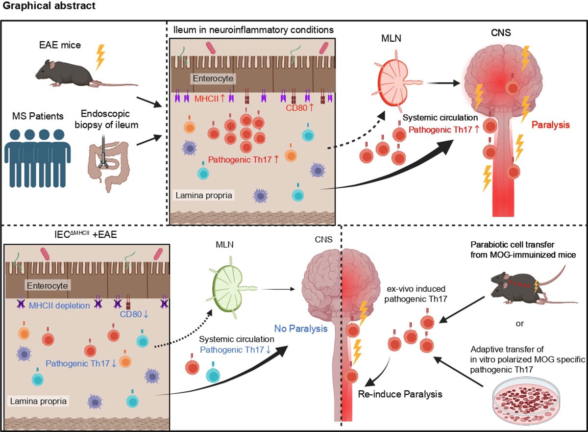
Dr. Suzuki, Dr. Sujino and colleagues built on earlier evidence that mild intestinal (ileal) inflammation appears in experimental autoimmune encephalomyelitis (EAE), a mouse model of MS. They then examined whether similar inflammation is also found in people with MS. Using single cell RNA sequencing on intestinal biopsies, the analysis showed that inflammatory Th17 cells accumulate both in the EAE mouse model and in the intestines of patients with MS, pointing to a conserved gut CNS axis that may operate in human disease.
In EAE mice and in patients with MS, intestinal epithelial cells (IECs) showed increased activity in antigen presentation pathways. Epithelial cells in the ileum had especially high expression of major histocompatibility complex class II (MHC II), which presents antigens to CD4+ T cells. When MHC II was selectively deleted in IECs, pathogenic Th17 cell generation and disease severity were reduced.
IECs usually do not present antigens to immune cells. To test whether they could perform this function, Dr. Suzuki, Dr. Sujino, and colleagues carried out co-culture assays. The findings showed that IECs can directly present antigens through an MHC II-dependent process and prime CD4+ T cells in the gut. In those assays, IECs also pushed activated CD4+ T cells toward Th17 polarization. The results made clear that the gut can serve as an important site where pathogenic CD4+ T cells are activated and become pro-inflammatory Th17 cells.
Gut cells reach the spine
To find out whether these Th17 cells directly add to the autoreactive cell population in the CNS, Dr. Suzuki, Dr. Sujino, and colleagues used transgenic mice that express the Kaede protein, which undergoes photoconversion from green to red fluorescence upon exposure to violet light. This system made it possible to precisely follow pathogenic Th17 cells that were induced in the intestinal lamina propria and then moved to the spinal cord, where they drove neuroinflammation.
Together, the study shows that MHC II expressed by IECs plays a critical role in expanding pathogenic Th17 cells that later migrate to the CNS during EAE. The findings provide a cellular mechanism linking gut immune responses to autoimmune neuroinflammatory disease. The work also shows that although systemic circulation allows T cells to move among immune tissues, interactions between epithelial and immune cells in the gut mucosa can strongly influence effector T cell responses in the brain.
“While current therapies for MS often target B cells, our study highlights the gut as an important therapeutic site. Modulating intestinal microbiota or antigen-presenting activity of IECs represents new approaches to treating autoimmune neurological diseases,” explained Dr. Suzuki, emphasizing the therapeutic implications of their findings.
A deeper understanding of immune activity in the gut mucosa could support the development of better treatments for disabling neurological diseases such as MS.
Reference: “Intestinal epithelial MHC class II induces encephalitogenic CD4 T cells and initiates central nervous system autoimmunity” by Shohei Suzuki, Kentaro Miyamoto, Anna Tojo, Yusuke Yoshimatsu, Toshiaki Teratani, Hitoshi Uchida, Yasuhiro Nemoto, Ryuichi Okamoto, Andreas Michael Sihombing, Toshiro Sato, Jin Nakahara, Takanori Kanai and Tomohisa Sujino, 27 March 2026, Science Immunology.
DOI: 10.1126/sciimmunol.aec1627
This work was supported by the Japan Science and Technology Agency (JST) through the Fusion Oriented Research for Disruptive Science and Technology (FOREST) program (grant number: 21457195); Grants-in-Aid from the Japan Society for the Promotion of Science (JSPS) (grant numbers: 20H00536, 20H03665, 21K18272, 23H02899, 23K27590, and 25K22627); the Japan Agency for Medical Research and Development (CREST grant number: 21gm1510002h0001); KGRI Challenge grant; Sakaguchi Memorial Foundation; and Miyarisan Pharmaceuticals.
Never miss a breakthrough: Join the SciTechDaily newsletter.
Follow us on Google and Google News.