
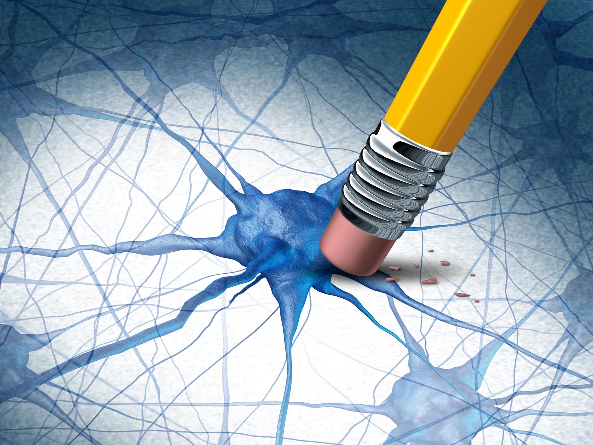
Alzheimer’s and stroke may begin at the brain’s outer defenses—not within neurons. A new study reveals how immune and vascular cells at the blood-brain barrier play a surprising and pivotal role in disease risk.
The Brain’s Hidden Defenders
The brain’s well-being depends on more than just neurons. It also relies on a specialized support system made up of blood vessels and immune cells. These cells act as protectors, deciding what substances can enter the brain, clearing out waste, and shielding it from potential harm by forming a structure called the blood-brain barrier.
Now, a new study led by scientists at Gladstone Institutes and UC San Francisco (UCSF) reveals that many of the genetic risk factors linked to neurological disorders such as Alzheimer’s and stroke appear to act within these protective cells.
“When studying diseases affecting the brain, most research has focused on its resident neurons,” says Gladstone Investigator Andrew Yang, PhD, senior author of the new study. “I hope our findings lead to more interest in the cells forming the brain’s borders, which might actually take center stage in diseases like Alzheimer’s.”
Published in the journal Neuron, the study offers answers to a long-standing question about where genetic risk actually begins. It suggests that weaknesses in the brain’s defense system may play a central role in the development of disease.
Decoding “Junk DNA”
For years, genetic studies have uncovered dozens of DNA variations that increase the likelihood of brain disorders like Alzheimer’s, Parkinson’s, and multiple sclerosis.
But one big question has remained: why do more than 90 percent of these DNA variants lie outside actual genes? These stretches of DNA, once labeled “junk,” do not directly create proteins. Instead, they help regulate gene activity, acting more like dimmer switches than lightbulbs.
Until recently, researchers lacked a comprehensive map showing how these regulatory regions affect specific genes and which brain cell types they influence. That gap has made it difficult to turn genetic insights into new treatments.

Breakthrough with MultiVINE-seq
The blood-brain barrier is the brain’s frontline defense—a cellular border made up of blood vessel cells, immune cells, and other supporting cells that meticulously controls access to the brain. Yet, these important cells have been difficult to study, even using the field’s most powerful genetic techniques.
To overcome this, the team at Gladstone developed MultiVINE-seq, a technology that gently isolates the vascular and immune cells from postmortem human brain tissue.
This technology allowed the team, for the first time, to simultaneously map two layers of information: the gene activity and the “dimmer switch” settings—known as chromatin accessibility—within each cell. The scientists studied 30 brain samples from individuals with and without neurological disease, giving them a detailed look at how genetic risk variants function across all major brain cell types.
Working closely with Gladstone Investigators Ryan Corces, PhD, and Katie Pollard, PhD, lead authors Madigan Reid, PhD, and Shreya Menon integrated their single-cell atlas with large-scale genetic data from studies of Alzheimer’s, stroke, and other brain diseases. This revealed where disease-associated variants are active—and many were found to be active in vascular and immune cells rather than neurons.
Revealing Cell-Specific Triggers
“Before this, we knew these genetic variants increased disease risk, but we didn’t know where or how they acted in the context of brain barrier cell types,” Reid says. “Our study shows that many of the variants are actually functioning in blood vessels and immune cells in the brain.”
One of the study’s most striking findings is that genetic risk variants affect the brain’s barrier system in fundamentally different ways, depending on the disease.
“We were surprised to see that the genetic drivers for stroke and Alzheimer’s had such distinct effects, even though they both involve the brain’s blood vessels,” Reid says. “That tells us they involve really distinct mechanisms: structural weakening in stroke, and dysfunctional immune signaling in Alzheimer’s.”
In stroke, genetic variants primarily affected genes responsible for the structural integrity of blood vessels, potentially weakening the vessels’ physical structure. Whereas in Alzheimer’s, the variants amplified genes that regulate immune activity, suggesting that overactive inflammation—not structural weakness—is the key issue.
PTK2B: The Immune Amplifier
Among the Alzheimer’s-associated variants, one stood out. A common variant near the PTK2B gene, which is found in more than a third of the population, was most active in T cells, a type of immune cell. The variant enhances expression of the gene, which may promote T cell activation and entry into the brain, putting immune cells into overdrive. The team found these super-charged immune cells near amyloid plaques, the sticky protein buildups that mark Alzheimer’s.
“Scientists are debating the role of T cells and related components of the immune system in Alzheimer’s,” Yang says. “Here, we provide genetic evidence in humans that a common Alzheimer’s risk factor may work through T cells.”
Excitingly, PTK2B is a known “druggable” target, and therapies that inhibit its function are already in clinical trials for cancer. The new study opens a fresh avenue to investigate whether such drugs could be repurposed for Alzheimer’s disease.
New Frontiers in Brain Protection
The study’s findings on the brain’s “guardian” cells point to two new opportunities for protecting the brain.
Located at the critical interface between the brain and the body, these cells are continually influenced by lifestyle and environmental exposures, which could synergize with genetic predispositions to drive disease. Their location also makes them a promising target for future therapies, potentially allowing for drugs that can bolster the brain’s defenses from the “outside” without needing to cross the formidable blood-brain barrier.
“This work brings the brain’s vascular and immune cells into the spotlight,” Yang says. “Given their unique location and role in establishing the brain’s relationship with the body and outside world, our work could inform new, more accessible drug targets and lifestyle interventions to protect the brain from the outside in.”
Reference: “Human brain vascular multi-omics elucidates disease-risk associations” by Madigan M. Reid, Shreya Menon, Hao Liu, Haoyue Zhou, Zhirui Hu, Simon Frerich, Bella Ding, Shahram Oveisgharan, Zimo Zhang, Sophia Nelson, Amanda Apolonio, David A. Bennett, Martin Dichgans, Katherine S. Pollard, M. Ryan Corces and Andrew C. Yang, 28 July 2025, Neuron.
DOI: 10.1016/j.neuron.2025.07.001
In addition to Yang, Reid, Corces, and Pollard, the study’s other authors are Shreya Menon, Hao Liu, Haoyue Zhou, Zhirui Hu, Bella Ding, Zimo Zhang, Sophia Nelson, and Amanda Apolonio of Gladstone; Simon Frerich of UC San Francisco; Shahram Oveisgharan and David A. Bennett of Rush University Medical Center; and Martin Dichgans of LMU Munich.
The work was supported by the National Institute of Neurological Disorders and Stroke (1R01NS128909-01), Alzheimer’s Association (ADSF-24-1345199-C, AARF-22-923641), BrightFocus Foundation (A2022027F), Cure Alzheimer’s Fund, the Ludwig Family Foundation, the Dolby Family Fund, the Bakar Aging Research Institute, National Institute of Mental Health (R01- 503 MH123178), National Institute of Aging (P01-AG073082, U01-AG072573), The Leducq Foundation (22CVD01, BRENDA), the Joachim Herz Foundation, and the National Human Genome Research Institute (UM1-HG012076).
Never miss a breakthrough: Join the SciTechDaily newsletter.
Follow us on Google and Google News.