Tiny lab-grown brain models and the particles they release may reveal hidden differences among Alzheimer’s patients.
Personalized treatment remains one of the biggest challenges in Alzheimer’s disease. Two patients can receive the same medication for symptoms such as depression, anxiety, or agitation and experience very different outcomes, leaving doctors with few ways to predict who will benefit.
Now, researchers at Johns Hopkins Medicine have developed miniature brain models grown from patients’ own cells that may help explain those differences. The study suggests these lab-grown tissues can capture key biological features of Alzheimer’s disease and reveal how individual patients might respond to commonly prescribed medications.
The researchers also identified promising clues inside tiny particles released by the organoids. These particles, known as extracellular vesicles, carry molecular information that could eventually help scientists diagnose Alzheimer’s disease, track its progression, and distinguish between different forms of the disorder.
The findings were published in Alzheimer’s & Dementia: The Journal of the Alzheimer’s Association.
Mini Brain Models Reveal Hidden Differences
Alzheimer’s disease affects more than 7 million Americans and remains the most common cause of dementia. Although recent advances have produced treatments that target disease-related proteins in the brain, many patients still depend on medications designed to manage behavioral and psychological symptoms that often accompany the condition.
Those treatments do not work equally well for everyone.
“Our study suggests that large-scale, patient-derived brain organoids and the vesicles they secrete can help us stage Alzheimer’s disease, investigate the mechanisms that drive it and assess how patient subgroups may respond to different treatments,” says study leader Vasiliki Machairaki, Ph.D., associate professor of genetic medicine at the Johns Hopkins University School of Medicine.
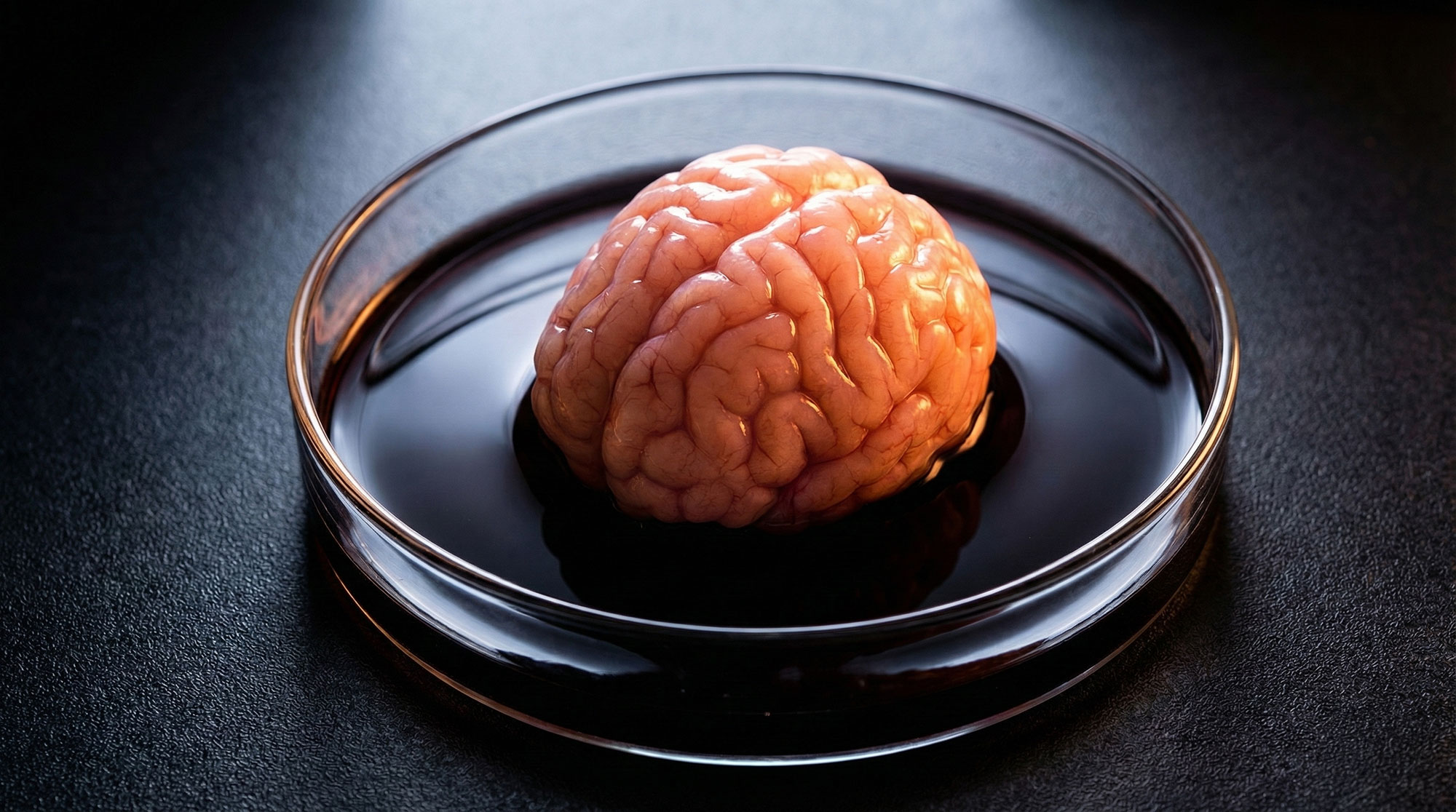
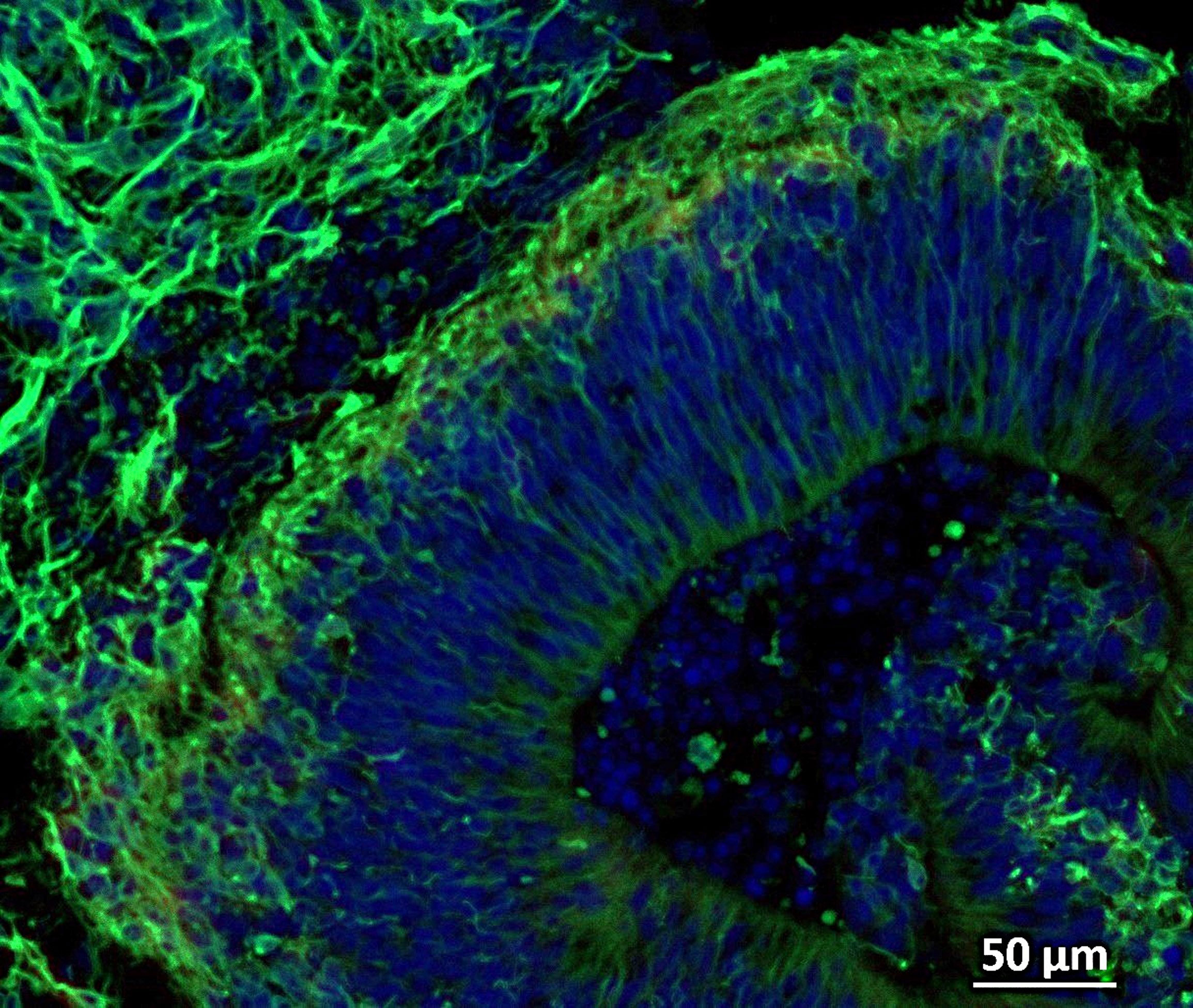
To investigate why patients respond differently, the team turned to brain organoids, small clusters of living brain tissue grown in the laboratory. Unlike conventional cell cultures, organoids can mimic some of the cellular organization and interactions found in the human brain, providing researchers with a more realistic model for studying disease.
The scientists began with blood samples collected from patients enrolled in the NIH-funded Johns Hopkins Alzheimer’s Disease Research Center. Those cells were reprogrammed into induced pluripotent stem cells, which can develop into nearly any cell type in the body.
Using stem cells from both Alzheimer’s patients and healthy volunteers, the researchers produced hindbrain organoids containing serotonin-producing neurons. The hindbrain plays an important role in regulating functions such as sleep, breathing, and heart rate, while serotonin signaling has long been linked to mood and behavior.
The cells organized themselves into pea-sized structures that resembled aspects of the human hindbrain. According to Machairaki, the study may be among the largest Alzheimer’s investigations to use patient-derived brain organoids, involving hundreds of organoids from both patients and healthy individuals.
Testing a Common Alzheimer’s Symptom Treatment
When the researchers compared organoids from Alzheimer’s patients with those from healthy individuals, they found clear molecular differences. Proteins involved in cell communication, inflammation, and other disease-related processes were altered in the Alzheimer’s-derived tissues.
The team then exposed the organoids to escitalopram oxalate, a commonly prescribed antidepressant in the selective serotonin reuptake inhibitor (SSRI) class.
Some organoids responded strongly, showing increases in proteins associated with serotonin signaling and communication between neurons. Others showed little or no measurable change.
That variation closely mirrors what doctors observe in real patients, where responses to the same medication can differ dramatically despite similar diagnoses.
“We used these organoids to model how some patients’ tissue may respond to a commonly prescribed SSRI,” Machairaki says. “On a large-scale level, our model may eventually be used to identify subgroups of patients, based on underlying molecular mechanisms, who are more likely to respond to certain drugs and thus help us to create precise, targeted treatments in the long run.”
Tiny Biological Messengers May Offer New Clues
The researchers also examined extracellular vesicles, microscopic particles released by cells that transport proteins and other biological material throughout the body.
Because these particles can be detected in bodily fluids such as blood, scientists are increasingly studying them as potential sources of disease biomarkers that could be measured without invasive procedures.
Before and after escitalopram treatment, the team analyzed extracellular vesicles released by both Alzheimer’s-derived organoids and healthy controls.
The vesicles contained proteins involved in memory, neurotransmitter release, and communication between neurons. Several proteins linked to Alzheimer’s disease, including RAB3A, NSF, and ATCAY, appeared at lower levels in organoids derived from patients with the disease.
After treatment, some of those proteins increased in certain samples, particularly proteins associated with serotonin signaling and synaptic activity.
As with the organoids themselves, responses varied considerably between samples. Some showed substantial molecular changes, while others showed almost none.
Toward More Personalized Alzheimer’s Care
The findings suggest that extracellular vesicles could eventually help identify which patients are most likely to benefit from specific therapies, potentially allowing treatments to be matched more closely to the biology of an individual’s disease.
The researchers are now working to create more advanced organoids that include immune cells and vessel-like networks that mimic blood vessels. These additions could make the models more representative of the living human brain and improve their value for studying Alzheimer’s disease.
Looking ahead, Machairaki envisions a future in which extracellular vesicles serve as a type of liquid biopsy, helping doctors diagnose Alzheimer’s disease, determine its stage, and identify distinct biological subtypes that may require different treatment approaches.
Reference: “Proteomic profiling of brain organoids and extracellular vesicles identifies early Alzheimer’s disease biomarkers and drug response heterogeneity” by Rachel J. Boyd, Daiyun Dong, Ram Sagar, Anton Iliuk, Waqar Ahmed, Xenia Androni, Anton P. Porsteinsson, Paul B. Rosenberg, Constantine G. Lyketsos, Kenneth W. Witwer and Vasiliki Mahairaki, 8 April 2026, Alzheimer’s & Dementia.
DOI: 10.1002/alz.71273
In addition to Machairaki, scientists who contributed to this work include Rachel Boyd, Daiyun Dong, Ram Sagar, Waqar Ahmed, Xenia Androni, Paul Rosenberg, Constantine Lyketsos and Kenneth Witwer from Johns Hopkins, Anton Iliuk from Tymora Analytical Operations and Anton Porsteinsson from University of Rochester School of Medicine and Dentistry.
Funding for this study was provided by the National Institutes of Health (T32 AG058527, R01AG052510, P30AG066507, 1RF1AG083801, AGR01054771, AGR01050515, AGR01046543 and AGR01071522), the Paul G. Allen Frontiers Foundation and the Richman Family Precision Medicine Center of Excellence in Alzheimer’s Disease at The Johns Hopkins University.
Never miss a breakthrough: Join the SciTechDaily newsletter.
Follow us on Google and Google News.