

Clearing amyloid plaques may break the chain reaction that fuels brain cell destruction in Alzheimer’s disease.
One Alzheimer’s brain offered researchers something they almost never get to see: a natural side-by-side experiment.
In some regions, years of anti-amyloid treatment had removed most of the sticky plaques associated with Alzheimer’s disease (AD). Those areas contained little or no tau, the abnormal protein closely tied to neuron loss and cognitive decline. Just millimeters away, neighboring regions where amyloid remained were packed with more tau, inflammation, and signs of continued brain damage.
The unusual pattern provides rare human evidence that removing amyloid may do more than reduce plaques. It may also interrupt the chain of biological events that allows Alzheimer’s to spread through the brain.
Researchers from the Perelman School of Medicine at the University of Pennsylvania presented the findings at the 2026 Alzheimer’s Association International Conference. The report was also published in JAMA.
“Seeing both disease patterns side by side in the same brain gave us a rare opportunity to understand how amyloid removal affects other proteins that contribute to Alzheimer’s disease,” said co-senior author David Wolk, MD, co-director of the Penn Memory Center and director of the Penn Alzheimer’s Disease Research Center. “The findings provide some of the clearest human evidence to date that anti-amyloid therapies may limit the accumulation of tau and slow the brain changes that lead to memory loss and cognitive decline.”
Why Amyloid and Tau Matter
Alzheimer’s is defined by two major protein abnormalities. Beta-amyloid collects outside neurons and forms plaques, while tau accumulates inside the cells and twists into tangles. Amyloid is thought to appear first, but tau is more closely associated with where neurons are dying and where symptoms are worsening.
That distinction has fueled a central debate in Alzheimer’s research over whether removing amyloid also slows the rest of the disease process.
Clinical trials can track memory, brain scans, and biological markers, but they cannot usually show exactly what happened inside individual brain regions over many years. Autopsy studies can provide that level of detail, yet very few people treated with anti-amyloid drugs have donated their brains for examination.
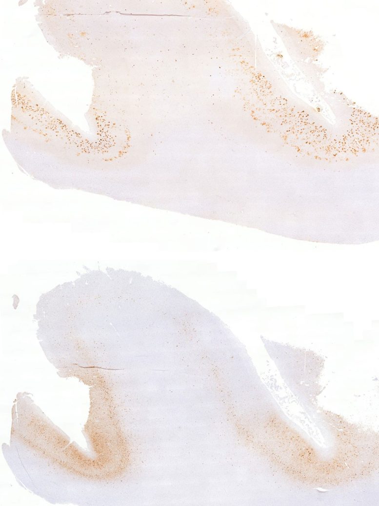
The new report centers on a man in his fifties who had mild cognitive impairment caused by AD. He received 30 doses of aducanumab during a clinical trial over four and a half years. He died about four years after his final dose and more than a decade after his symptoms began.
Before his death, he chose to donate his brain to the Center for Neurodegenerative Disease Research within the Department of Pathology and Laboratory Medicine.
A Patchwork Response to Treatment
The examination revealed a patchwork rather than a uniform response.
The gyri, the raised ridges of the brain’s folds, had lost most of their amyloid and showed very little tau. In the nearby sulci, the grooves between those ridges, amyloid remained abundant. Those regions also had far more tau, inflammation, and neurodegeneration.
Brain scans collected while the participant was alive had already shown that the treatment cleared amyloid unevenly. The autopsy allowed researchers to connect that imaging pattern with the later distribution of tau and brain damage.
“Many previous reports have shown either extensive amyloid clearance or limited clearance,” said co-senior author Edward B. Lee, MD, PhD, the John Q. Trojanowski, MD, PhD Professor for Neurodegenerative Disease Research in Pathology and Laboratory Medicine. “This case represented a unique ‘Goldilocks’ scenario in which some regions cleared amyloid and others did not. It allowed us to directly compare what happened next in neighboring brain regions and better understand the relationship between amyloid, tau, and neurodegeneration.”
What the Findings Could Mean for New Drugs
Anti-amyloid drugs have rapidly changed the treatment of early Alzheimer’s. Aducanumab became the first therapy in the class to receive accelerated FDA approval, although it is no longer marketed. Newer drugs, including donanemab (Kisunla) and lecanemab (Leqembi), use a similar strategy and have received full FDA approval.
The findings therefore have relevance beyond a single experimental treatment. They suggest that the degree and location of amyloid removal may influence what happens later to tau and the surrounding brain tissue.
The Limits of a Single Case
However, the report describes only one patient. It cannot prove that amyloid clearance caused the regional differences or establish how much plaque must be removed to protect neurons.
It also raises a timing question. Most major treatment trials follow patients for about 18 months, but the biological effects seen in this brain unfolded over several years. Some benefits of amyloid removal may therefore take longer to become visible, especially if the treatment slows a disease process that has been developing silently for decades.
Researchers now want to understand why the drug cleared plaques more effectively from some brain regions than others. Identifying the biological pathways behind that uneven response could help scientists improve future treatments.
Could Earlier Treatment Prevent Brain Damage?
Another priority is determining whether therapy works best before tau has spread widely. Amyloid can build up long before memory problems appear, creating a potential window in which treatment might prevent or delay the damage that eventually produces symptoms.
“Patients may accumulate amyloid for years before symptoms appear,” said first author Christopher A. Brown, MD, PhD, an assistant professor of neurology. “This case suggests that removing amyloid early may help limit the changes that ultimately damage brain cells. Ongoing trials will help determine whether starting treatment before symptoms begin can provide even greater benefit.”
Reference: “Clinicopathologic Evaluation of Amyloid Clearance in Alzheimer Disease” by Christopher A. Brown, John L. Robinson, Sandhitsu R. Das, Emily McGrew, EunRan Suh, Viviana M. Van Deerlin, David J. Irwin, Ilya M. Nasrallah, Dawn Mechanic-Hamilton, Paul A. Yushkevich, David A. Wolk and Edward B. Lee, 12 July 2026, JAMA.
DOI: 10.1001/jama.2026.13058
This research was supported by the National Institutes of Health (P30-AG072979, RF1-AG069474, R01-AG056014, R01-AG055005, R01-AG072796, R01-AG065341, P01-AG084497).
Never miss a breakthrough: Join the SciTechDaily newsletter.
Follow us on Google and Google News.