
The study reveals the biology behind why the disease appeared so different after spreading to cows and offers a framework for recognizing new forms of the disease more quickly.
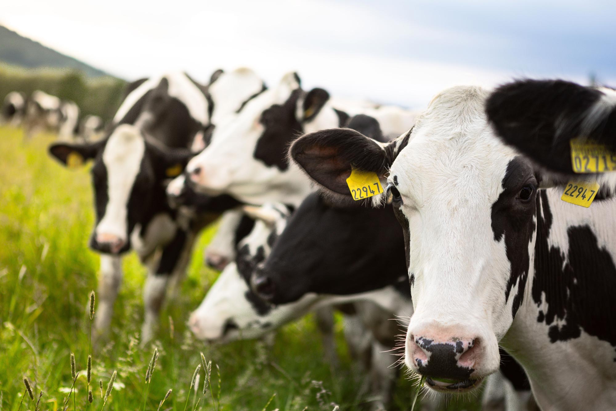
H5N1 bird flu caused severe infections in cows’ udders while largely sparing their lungs when it began spreading among U.S. dairy cattle in early 2024. That pattern made the disease difficult to recognize. In other mammalian species, H5N1 affects the lungs.
Researchers at the University of Pittsburgh School of Public Health have now traced that difference to the molecular structures the virus uses to attach to cells. The structures that allowed H5N1 to bind were abundant in udder tissue but nearly absent from cows’ airways. Published in Science Advances, the study provides the first explanation of the biological mechanism behind this pattern.
Why H5N1 favored cows’ udders
For influenza viruses, the ability to infect a particular animal or organ depends partly on finding compatible receptors on its cells. These receptors include certain glycans, sugar-based molecules that interact with the virus much like a lock and key.
Suresh Kuchipudi, Ph.D., the study’s senior author and chair of Infectious Diseases and Microbiology at Pitt Public Health, has spent his career studying influenza, with a particular focus on how receptor biology shapes infection. But standard methods for detecting receptors had left an unresolved question in cattle.
Earlier studies by other groups used staining techniques to identify flu-related glycan receptors in cows’ noses, tracheas, and lungs. Their presence did not explain why the animals were largely escaping respiratory infection. The researchers needed to determine which of those receptors H5N1 could actually use.
“Glycan biology is very complex,” said Kuchipudi. “We realized that, to understand what was really going on, we would need to use more innovative technologies and map out the fine‑detailed architecture that enables the virus to bind to cells.”
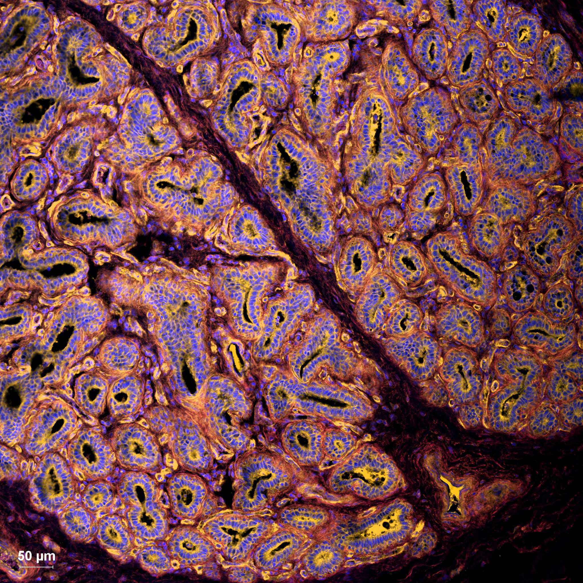
Kuchipudi collaborated with Lauren E. Pepi, Ph.D., of Harvard Medical School, an expert in glycomics, the comprehensive cataloging of glycan structures. The team combined experiments that tested viral binding with staining methods and ultra-high-resolution imaging to examine the receptors in greater detail.
In those experiments, only a particular subtype, called N-linked sialic acid receptors, could bind to H5N1. Their distribution explained the contrast between the airways and udders. These receptors were virtually absent from cow airway tissue but widespread in udders, making them a “perfect breeding ground for the virus,” Kuchipudi said.
Udder infections delayed H5N1’s detection
That preference for udder tissue helps explain the difficulty veterinarians faced when the disease first appeared in dairy cattle along the Texas Panhandle. The animals developed persistent cases of severe, necrotizing mastitis, a painful inflammation that damages tissue in the mammary glands, where milk is produced.
“Mastitis is a classic disease in milk-production animals, and veterinarians were dutifully looking to all the usual suspects for the source, like bacterial pathogens,” said Kuchipudi. “When the real culprit turned out to be bird flu, everyone in the field was caught completely by surprise. We hadn’t even remotely considered that cattle could be a host for H5N1.”

During the weeks before H5N1 was identified, the virus spread between herds, sickening cattle and contaminating their surroundings. Infection in the udders also created a route for the virus to leave the animals through their milk.
“If a cow is infected, it sheds a lot of virus into the milk,” said Kuchipudi. “This raised concerns about occupational risk for farm workers. Also, there is a habit of feeding raw milk to domestic pets, like cats, and there have been instances of cats dying, which we studied previously.”
Pasteurization effectively kills the virus, Kuchipudi emphasized, underscoring the importance of avoiding raw milk.
Receptors could flag other vulnerable hosts
H5N1 now affects more than 100 bird and mammal species worldwide. The researchers propose using their approach to assess whether other species could become hosts and which tissues might be vulnerable. Recognizing an unfamiliar pattern of infection sooner could give public health officials more time to take measures to limit its spread.
“We can preemptively screen different species and different tissues within them for susceptibility,” said Kuchipudi. “For example, would they exhibit respiratory symptoms? Would they show only mastitis, as in cows? Or would they show neurological disease, as our team has shown in cats? The lessons learned could potentially help prevent us from being caught by surprise again.”
Reference: “Receptor basis of unusual tissue tropism of avian influenza H5N1 clade 2.3.4.4b virus in cattle” by Surabhi Srinivas, Shubhada K. Chothe, Santhamani Ramasamy, Sougat Misra, Noel Chandan Nallipogu, Lindsey LaBella, Yin-Ting Yeh, May Wang, Lauren E. Pepi, Heidi L. Pecoraro, Brett T. Webb and Suresh V. Kuchipudi, 19 June 2026, Science Advances.
DOI: 10.1126/sciadv.aea2068
This research was supported by Pitt Public Health, and the U.S. Department of Agriculture’s National Institute of Food and Agriculture (FP00039373/AWD00010780).
Never miss a breakthrough: Join the SciTechDaily newsletter.
Follow us on Google and Google News.