Researchers at Trinity College Dublin, working together with the Royal College of Surgeons in Ireland (RCSI), have developed special fluorescent, color-changing dyes that, for the first time, can be used to simultaneously visualize multiple distinct biological environments using only one singular dye.
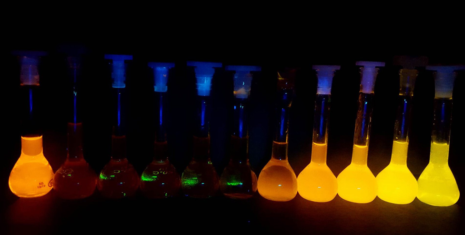
When these dyes are encapsulated in delivery vessels, like those used in technologies like the COVID-19 vaccines, they “switch on” and give out light via a process called “aggregation-induced emission” (AIE). Soon after delivery into the cells their light “switches off” before “switching on” again once the cells shuttle the dyes into cellular lipid droplets.
Advanced Imaging Techniques
Because the light coming from inside the cells is of a different color and occurs within a different time window to the light coming from the same dye inside the delivery vessels, the researchers can use a technique called “fluorescence lifetime imaging” (FLIM) to distinguish between the two environments in real-time.
The work was recently published in the leading international journal, Chem. First author, Dr Adam Henwood, Senior Research Fellow in the School of Chemistry and based at the Trinity Biomedical Sciences Institute (TBSI), worked on this design with PhD student Connie Sigurvinsson.
Dr Henwood explained: “Bioimaging relies on “on/off” dyes where the dyes only emit light under one set of conditions but are otherwise switched off. This is extremely useful, but it does mean that you can only look at one place at a time under your microscope. The exciting part about this work is that our dyes hit a sweet spot that gives them distinctive on/off/on properties and, crucially, we can both observe and differentiate these different “on” states.
“So, we both see more and see better than before. We do this by timing how long it takes for the light coming from our samples to reach the microscope: light from the delivery vessels takes marginally more time than light from within the cells. By collecting enough light signals, we can use this information to rapidly build up precise 3D images of the two different dye environments. The time differences are small – just a few billionths of a second either way – but our method is sensitive enough to capture it.”
This unique quality means the dyes could have a huge suite of applications and, for example, hold the potential to revolutionize bio-sensing and imaging approaches.
Because these dyes can help scientists map the intricate structures within living cells with such high contrast and specificity, they could help illuminate how drugs are taken up and metabolized by cells or allow scientists to design and conduct a range of new experiments to better our understanding of the complex inner workings of cells and their all-important biochemical machinery.
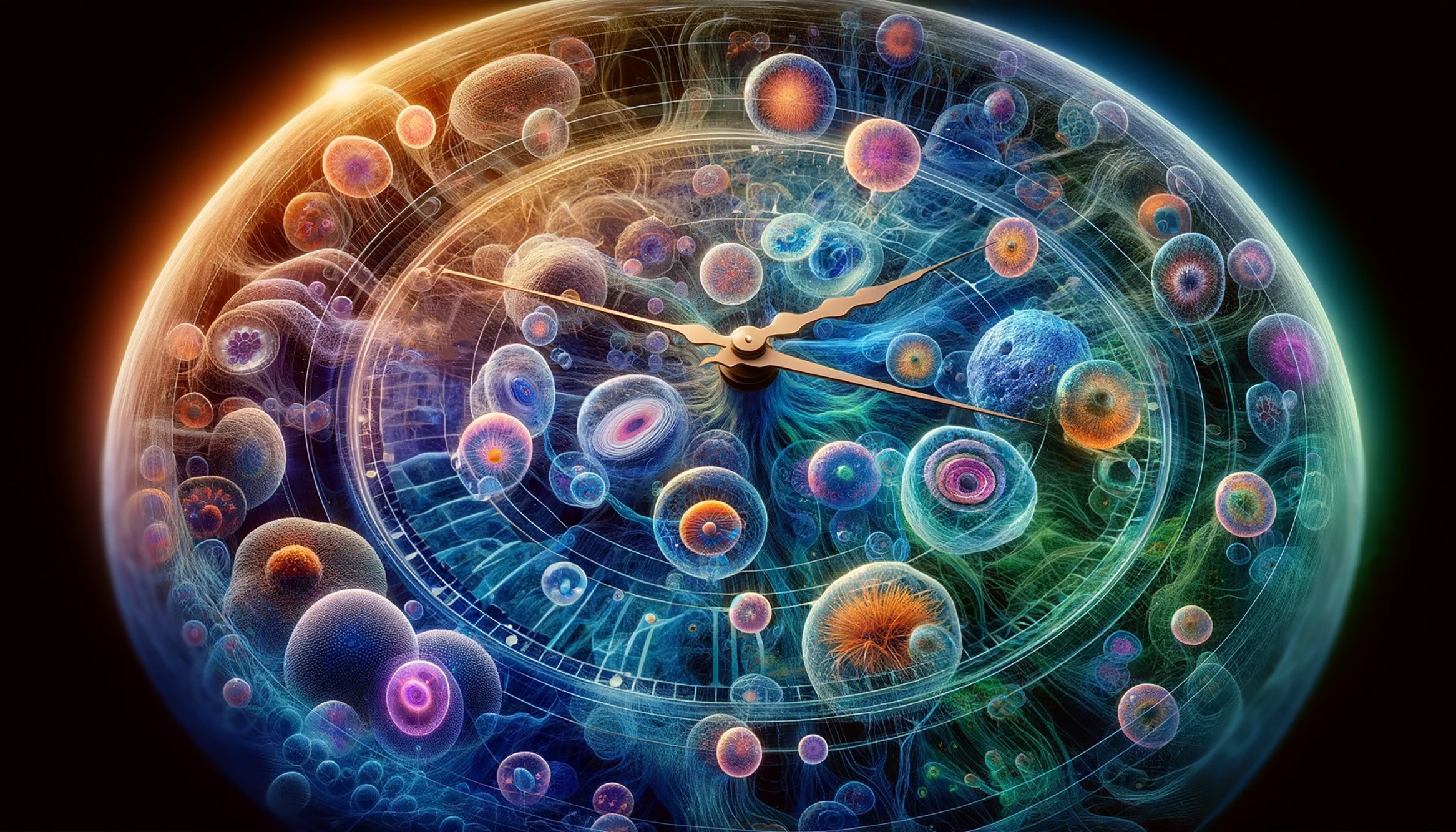
In the published journal article, the scientists focused on using the dyes to image cellular lipid (fat) droplets, which are one example of important “organelles” that make up living cells in most complex organisms (like us humans).
Lipid droplets, once considered to be simple “fat reservoirs”, are now believed to play an important role in regulating cellular metabolism, coordinating lipid uptake, distribution, storage, and use in the cells. Because of this growing understanding of their importance, and because sudden changes in their activity often indicate cellular stress, they serve as a useful test case scenario for the dyes. One potential avenue of further research is to see whether the team can target other important cellular organelles with their dyes.
Thorfinnur Gunnlaugsson, Professor of Chemistry in the School of Chemistry at Trinity and based in TBSI, is the senior author of the article. He said:
“Being able to monitor cellular function or the flow of molecules or drug candidates within cells by observing different fluorescence emission colors is extremely attractive. The breakthrough here is that we can resolve and use the difference in their fluorescence lifetimes to identify these same probes within different cellular environments in a fast and accurate manner, literally allowing us to map out their colorful “time travel” within the cells.
“Most exciting, however, is that this phenomenon does not apply to cellular imaging. These results open up new possibilities in everything from studying chemical biology, as we have shown here, to many other medical applications and even in the generation of novel functional materials for use beyond biology. Any molecular or nanomaterial that requires controlled molecular motion can in principle be mapped and fine-tuned using our new method.”
Potential Applications and Future Directions
And indeed, it is here where the authors intend to cast the net far and wide. They envisage many new possibilities for these dyes, pointing toward their exceptional sensitivity as attractive for developing sensors of hazardous environmental pollutants or using their bright, light-emitting properties to power chemical transformations, analogous to nature’s own photosynthesis.
The research has both an international (eight nations are represented) and Irish feel to it, with the latter’s key funding bodies the Irish Research Council (IRC) and Science Foundation Ireland both playing critical financial support roles. Most notable is SFI’s Research Centre for Pharmaceuticals, SSPC, which principally funded the work, as well as contributions from the SFI AMBER center and through the AMBER-based EPSRC-SFI Centre for Doctoral Training Programme.
Prof. Damien Thompson, Professor of Physics at the University of Limerick and Director of the SSPCsaid: “As a center, we keep pushing forward and creating new knowledge at the interface of materials and biology. This collaborative work between two of our principal investigators at Trinity and RCSI showcases the power of fundamental science to drive innovation in medicine. The closer we look at the molecule-cell interface, and crucially, the better we can see, in real-time, how molecules diffuse from place to place inside the cell nanomachinery, the closer we get to realising Richard Feynman’s dream of understanding everything that living things do from the wiggling and jiggling of atoms.
“But only recently have researchers had sufficient experimental and computational resources to track these motions and vibrations in complex biological environments. This exciting new work demonstrates more specific, high contrast imaging of subcellular dynamics, which will in turn enable researchers to develop more effective drug formulations with reduced side effects.”
Professor Donal O’Shea, who oversaw the investigation, is an expert in cell imaging based in RCSI’s Department of Chemistry and Super-Resolution Imaging Consortium (funded by Science Foundation Ireland, SFI). He added: “Our use of FLIM to track dynamic AIE interactions with living cells is an approach that can have broad applicability for other fluorophore systems allowing insights to be gained that were previously hidden.”
Reference: “Time-resolved fluorescence imaging with color-changing, “turn-on/turn-on” AIE nanoparticles” by Adam F. Henwood, Niamh Curtin, Sandra Estalayo-Adrián, Aramballi J. Savyasachi, Tómas A. Gudmundsson, June I. Lovitt, L. Constance Sigurvinsson, Hannah L. Dalton, Chris S. Hawes, Denis Jacquemin, Donal F. O’Shea and Thorfinnur Gunnlaugsson, 1 December 2023, Chem.
DOI: 10.1016/j.chempr.2023.10.001
The study was funded by the Irish Research Council and the Science Foundation Ireland.
Never miss a breakthrough: Join the SciTechDaily newsletter.
Follow us on Google and Google News.
3 Comments
Yeah
Azo Compounds? https://en.wikipedia.org/wiki/Azo_compound (When I first heard about these while taking Organic Chemistry, I thought about instant camo paint for military vehicles and aircraft, and color change paints in general, but never this.) Could these be useful in cancer detection? Or to instantly detect other diseases?
The headline is misleading
It’s enabling mapping of changes with time…..timelapse
Not time travel