
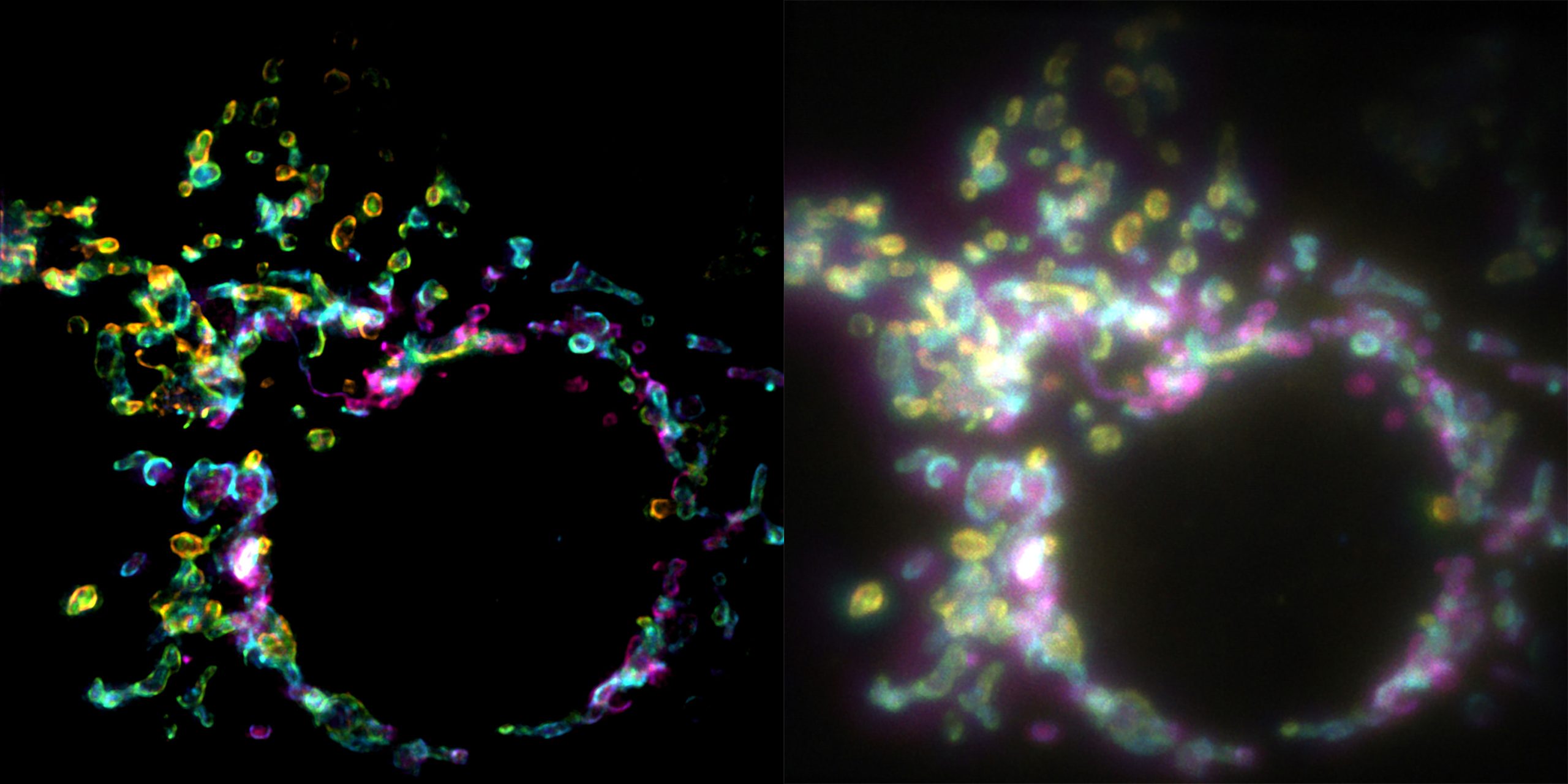
A new imaging technique allows scientists to capture fleeting cellular processes that conventional microscopes struggle to resolve.
Living cells are constantly in motion, but capturing their smallest structures in sharp detail has long been a challenge. Traditional super-resolution microscopy often requires hundreds or thousands of images to reveal features too small for conventional light microscopes. When structures move during that process, the resulting images can become blurred. Now, researchers have developed a technique that captures these microscopic details in a single camera exposure.
The new method, called Spatial Polarization-Induced Fluorescence Fluctuation Imaging (SPIFFI), was developed by a team led by Aleksandra Radenovic at the Laboratory of Nanoscale Biology (LBEN) in EPFL’s School of Engineering. Published in Nature Methods, the research demonstrates up to a twofold improvement in image resolution, allowing scientists to distinguish structures as small as 160 to 170 nanometers while observing living cells.
Why Moving Cells Are So Difficult to Image
Super-resolution microscopy has transformed cell biology by revealing structures that conventional light microscopes cannot clearly distinguish. Many established techniques rely on fluorescent molecules that emit light when illuminated by a laser. Scientists analyze subtle changes in this fluorescence across large sequences of images, then combine the information to reconstruct a sharper picture.
These approaches work particularly well with chemically preserved cells, where structures remain stationary during imaging. Living cells present a different challenge. Mitochondria continually change shape, microtubules reorganize, and DNA moves within the cell. Capturing multiple frames takes time, allowing these structures to shift before the final image can be assembled.
Video demonstrating the ability of SPIFFI to capture rapid cellular dynamics and extract molecular orientation at the same time. Credit: EPFL LBEN CC BY SA 4.0
“Essentially, previous approaches used temporal information to resolve spatial resolution, but this doesn’t work very well on living cells,” explains first author and LBEN PhD student Wei Guo.
Using Polarized Light to Reveal Hidden Structures
Instead of collecting information over many exposures, SPIFFI takes advantage of a property of fluorescent light known as polarization. Fluorescent molecules emit light whose waves tend to oscillate in particular directions, depending on the molecules’ orientation. These directional differences contain spatial information that conventional fluorescence imaging does not fully exploit.
SPIFFI separates the emitted light into four channels, each sensitive to a different polarization component. By comparing the four images captured during the same exposure, the system can recover fine structural details without needing to accumulate information across hundreds of frames.
The researchers demonstrated the technique by imaging moving mitochondria and microtubules, as well as cellular structures undergoing fusion and splitting. These rapid changes are difficult to resolve with methods that require multiple exposures to produce one super-resolution image.
“Our experiments show that SPIFFI can capture fast-moving processes within cells, while enabling high-throughput, multi-dimensional imaging beyond the limits of conventional microscopes,” Radenovic summarizes.
Turning Super-Resolution Images Into Videos
Because SPIFFI produces a super-resolved image from every camera exposure, successive frames can be combined to create videos of cellular activity without sacrificing spatial resolution through multi-frame reconstruction.
“With previous techniques, taking many images would only result in one super-resolved frame. With SPIFFI, every frame is super-resolved, meaning we can now produce super-resolution videos of live cells,” Guo says. “We also seamlessly integrated SPIFFI images with existing fluctuation-based methods for post-processing, achieving resolutions of about 80 nanometers.”
The team is now working to make the imaging system more compact and integrate it with three-dimensional microscopy. Because SPIFFI’s optical components can be incorporated into existing fluorescence microscopes, the technique could eventually support research in cell biology, neuroscience, biophysics, and drug discovery, particularly when scientists need to observe rapid structural changes inside living cells.
Reference: “SPIFFI enables single-shot super-resolution and multidimensional imaging” by Wei Guo (郭威), Lely Feletti and Aleksandra Radenovic, 31 August 2026, Nature Methods.
DOI: 10.1038/s41592-026-03196-6
Never miss a breakthrough: Join the SciTechDaily newsletter.
Follow us on Google and Google News.