Brain scans suggest long COVID’s biggest clues may lie in the brain’s emotion centers, not widespread inflammation.
A new brain imaging study suggests that persistent symptoms of long COVID may not be driven by ongoing brain inflammation, as many researchers have suspected.
Scientists in Finland used advanced imaging techniques to examine the brains of people experiencing long-lasting symptoms after COVID-19 infection. While long COVID has often been linked to lingering inflammation in the brain, the researchers found no evidence of widespread neuroinflammation when comparing patients with long COVID to healthy individuals.
“We did not observe evidence of widespread brain inflammation in patients with long COVID when compared to healthy controls,” says Professor of Neuroimmunology and InFLAMES Research Flagship group leader Laura Airas, who led the study.
Comparing Long COVID With Healthy People and MS Patients
The study involved 14 people with long COVID, 11 healthy volunteers, and 13 individuals with multiple sclerosis (MS), a neurological disease known to cause inflammation in the brain.
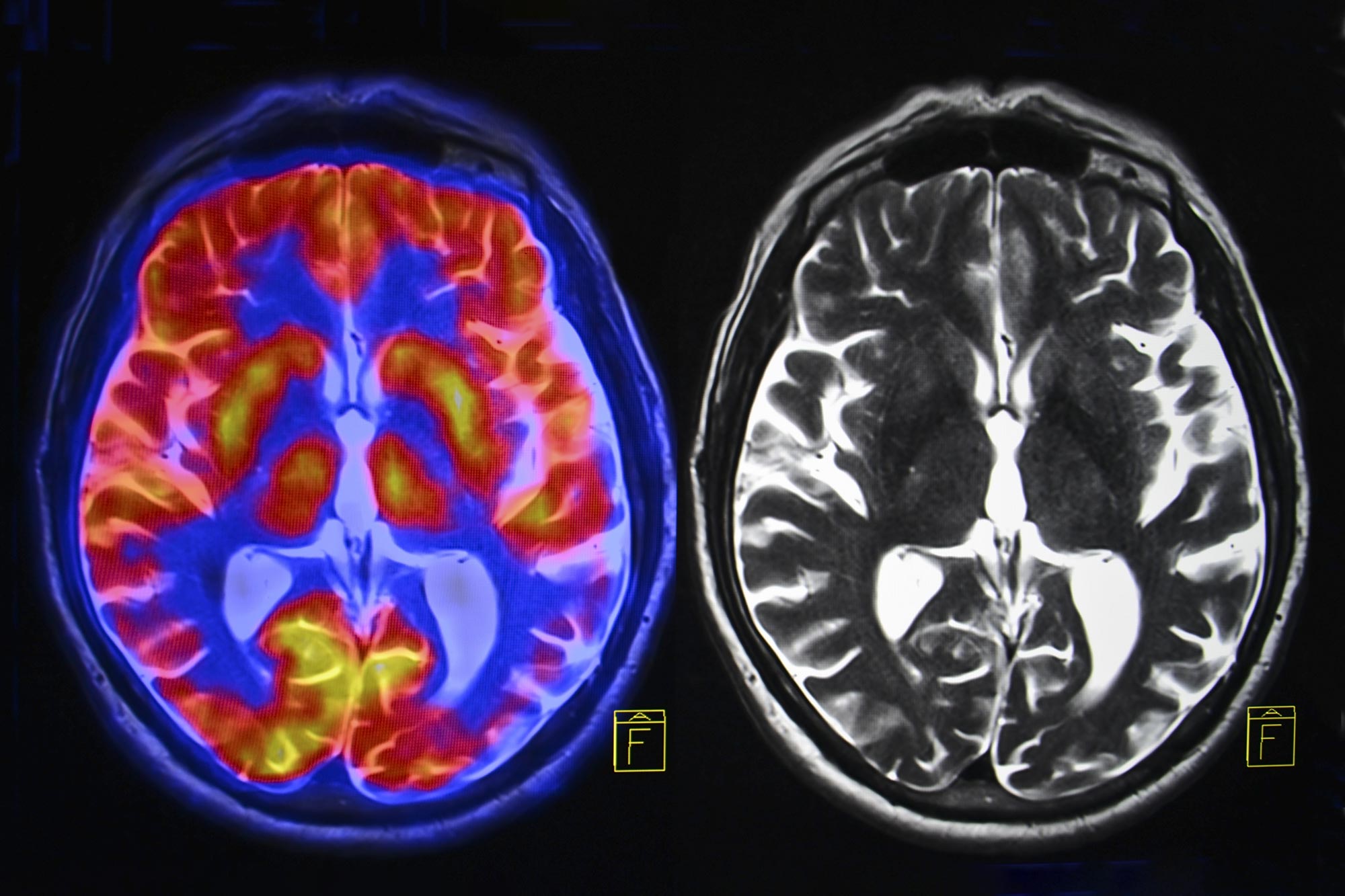
Researchers performed positron emission tomography (PET) scans designed to detect neuroinflammation, along with magnetic resonance imaging (MRI) scans to evaluate brain structure and white matter changes. Blood samples were also collected to measure biomarkers associated with damage to neurons and glial cells.
The results showed that people with long COVID had much lower inflammatory activity in the brain’s white matter than patients with MS. Researchers also found no differences between long COVID patients and healthy participants in markers associated with brain inflammation or neurodegeneration.
Signs of Inflammation May Fade Over Time
Previous neuropathological studies of severe acute COVID-19 have documented clear evidence of brain inflammation. However, the new findings suggest that any inflammatory effects linked to long COVID may be strongest earlier in the course of the illness.
Among participants with long COVID, those who underwent brain scans within 16 months of their infection showed higher levels of inflammatory activity in white matter than those who had been living with the condition for a longer period.
According to Airas, the findings indicate that inflammation may be more prominent during the earlier stages of the disease and gradually decline over time.
Emotional Brain Regions Linked to More Severe Symptoms
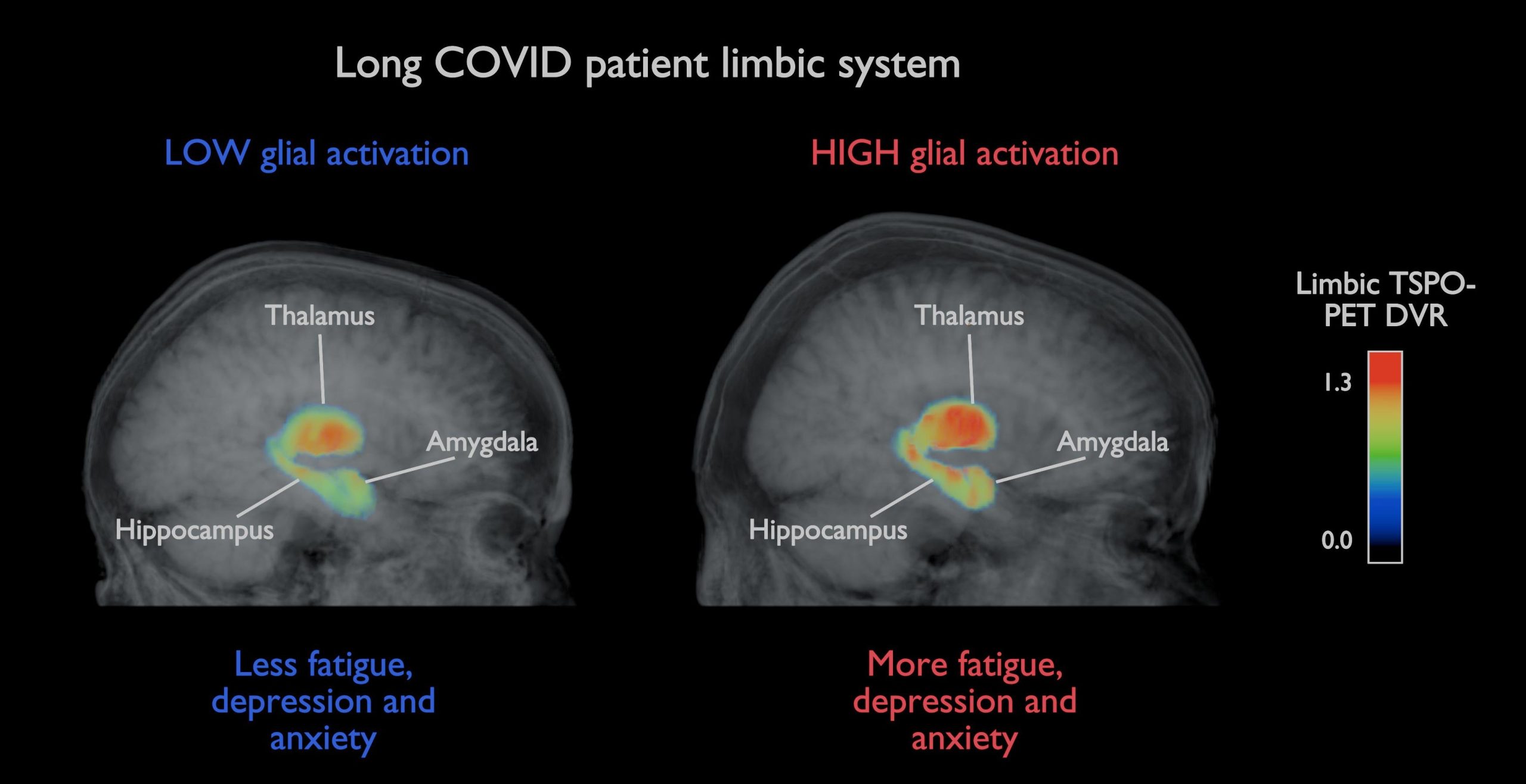
One of the study’s most notable findings involved two brain regions that help regulate emotions, memory, and responses to stress.
Researchers found that people reporting higher levels of anxiety and depression, as well as poorer quality of life, tended to show increased cellular activity in the hippocampus and amygdala.
These observations suggest that changes in activity within emotion-regulating areas of the brain may be connected to symptom severity in at least some people with long COVID.
What the Findings Could Mean for Future Treatments
The researchers say the results add to a growing understanding of long COVID and challenge the idea that persistent brain inflammation is the main cause of prolonged symptoms in all patients.
Instead, the study points to a more complicated picture. Inflammatory changes may occur soon after infection but appear to lessen over time, while other biological processes may contribute to symptoms that continue for months or even years.
Long COVID is a recognized condition affecting millions of people worldwide. Common symptoms can include fatigue, cognitive difficulties, anxiety, depression, and other health problems that persist long after the initial infection.
The findings also raise the possibility that some patients could benefit more from treatments focused on stress management and emotional regulation rather than therapies aimed solely at reducing inflammation.
“This study highlights the need to continue investigating the complex biological mechanisms underlying long COVID. Understanding these processes is essential for developing targeted treatments,” notes Airas.
The study by Airas and colleagues was published in the Journal of Neurology.
Reference: “Association between post-COVID-19 neuropsychiatric symptoms and persistent glial activation in the limbic system: a TSPO PET study” by Joel Tuomaala, Maija Saraste, Emma Smith, Matilda Kuusi, Elisabet Westerberg, Eveliina Honkonen, Rahim Kargar, Sini Laaksonen, Jussi Lehto, Amelie Luoma, Markus Matilainen, Olavi Misin, Janne Atosuo, Mari Kanerva, Helena Liira, Sini Laakso, Tatiana Posharina, Virva Saunavaara, Saara Wahlroos, Johan Rajander and Laura Airas, 30 April 2026, Journal of Neurology.
DOI: 10.1007/s00415-026-13842-w
InFLAMES Flagship is a joint initiative of the University of Turku and Åbo Akademi University, Finland. The program brings together immunology and related research fields to develop new diagnostic and treatment tools for personalized medicine. InFLAMES is part of the Research Council of Finland’s Flagship Program.
Never miss a breakthrough: Join the SciTechDaily newsletter.
Follow us on Google and Google News.
1 Comment
Howz the song go? “Don’t worry, Be happy – cause everythingz gonna be all right…”