The second week of gestation represents a critical stage of embryo development, or embryogenesis. Failure of development during this time is one of the major causes of early pregnancy loss.
Using surplus human embryos voluntarily donated from in vitro fertilization clinics in the UK, scientists have extensively studied the pre-implantation period in the laboratory, the period before a developing embryo would implant into the mother’s uterus on the seventh day after fertilization. Very little is known about the development of the human embryo after implantation occurs, however, as the embryo becomes inaccessible for study.
In 2016, Magdalena Zernicka-Goetz, now Caltech’s Bren Professor of Biology and Biological Engineering, and her team at the University of Cambridge developed a technique to culture human embryos outside the body of the mother beyond implantation. This enabled human embryos to be studied up until day 14 of development for the first time.
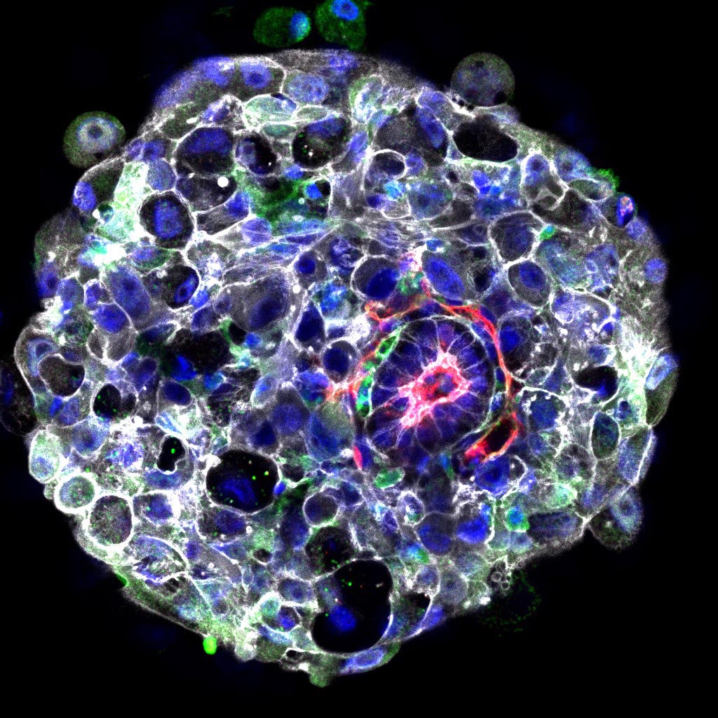
In a new study, researchers in the Zernicka-Goetz lab examined what happens at the molecular level during this early stage of embryogenesis. Their findings provide the first evidence that a disc-shaped group of cells located outside of the embryo known as the hypoblast sends a message to the embryo that initiates the formation of the head-to-tail body axis, which gives the previously symmetrical embryo two distinct ends, a head and a “tail.” The findings reveal that the molecular signals involved in the formation of the body axis in human embryos are similar to those in other mammals, despite significant differences in the positioning and organization of the cells of the embryos of different species.
The results are described in a paper appearing in the journal Nature Communications.
Molecular Messaging Between Embryo and Hypoblast
“We have revealed the patterns of gene expression in the developing embryo just after it implants in the womb, which reflect the multiple conversations going on between different cell types as the embryo develops through these early stages,” says Zernicka-Goetz. “We were looking for the genetic conversations that will allow the head to start developing in the embryo and found that these were initiated by cells in the hypoblast, which would not normally contribute to building the body itself. They send the message to the adjoining embryo cells, which respond by saying ‘OK, now we’ll set ourselves aside to develop into the head end.'”
The study identified these gene conversations in the developing embryo by sequencing the code in the thousands of messenger RNA (mRNA) molecules made by individual cells; mRNA molecules are translated by cellular machinery into the protein molecules that perform vital functions and give cells their structure. This allowed the researchers to capture changes in the evolving molecular profile of developing embryos after their implantation in the womb and revealed the embryonic cells’ progressive loss of pluripotency (the ability to give rise to any type of cell) as they developed into the distinct cell types that eventually give rise to all the organs of the human body.
A Window into Life’s Earliest Decisions
“Our goal has always been to enable insight to very early human embryo development in a dish to understand how our lives start. By combining our new technology of culturing human embryos with advanced sequencing methods, we have delved deeper into the key changes that take place at this incredible stage of human development when the embryo becomes remodeled to undertake its critical decisions at a time when so many pregnancies fail,” says Zernicka-Goetz.
For more on this research, see Key Molecular Events in the Developing Human Embryo Identified.
Reference: “A single cell characterisation of human embryogenesis identifies pluripotency transitions and putative anterior hypoblast centre” by Matteo A. Molè, Tim H. H. Coorens, Marta N. Shahbazi, Antonia Weberling, Bailey A. T. Weatherbee, Carlos W. Gantner, Carmen Sancho-Serra, Lucy Richardson, Abbie Drinkwater, Najma Syed, Stephanie Engley, Philip Snell, Leila Christie, Kay Elder, Alison Campbell, Simon Fishel, Sam Behjati, Roser Vento-Tormo and Magdalena Zernicka-Goetz, 17 June 2021, Nature Communications.
DOI: 10.1038/s41467-021-23758-w
Matteo Molè of the University of Cambridge is the study’s first author. In addition to Zernicka-Goetz, additional co-authors are Tim Coorens and Carmen Sancho-Serra of the Wellcome Sanger Institute; Marta Shahbazi, Antonia Weberling, Bailey Weatherbee, and Carlos Gantner in the Zernicka-Goetz laboratory of the University of Cambridge; embryologists Lucy Richardson, Abbie Drinkwater, Najma Syed, and Stephanie Engley of the Herts & Essex Fertility Centre at Bishops College; Philip Snell, Leila Christie, and Kay Elder of Bourn Hall in Cambridge, UK; Alison Campbell of CARE Fertility group in Nottingham, UK; Simon Fishel of CARE Fertility group and Liverpool John Moores University in Liverpool, UK; and finally, the collaborators responsible for analyses of sequencing data: Sam Behjati of the Wellcome Sanger Institute and the University of Cambridge; and Roser Vento-Tormo of the Wellcome Sanger Institute. Funding was provided by European Molecular Biology Organisation, the UKRI Medical Research Council, the Gates Cambridge Trust, the Wellcome Trust, and in the U.S. by Open Philanthropy (Silicon Valley), and the Curci and Weston Havens foundations.
Never miss a breakthrough: Join the SciTechDaily newsletter.
Follow us on Google and Google News.