Researchers have created synthetic mouse embryos from stem cells with beating hearts and brain foundations, providing insights into early pregnancy development.
Scientists have created model mouse embryos from stem cells that have beating hearts, as well as the foundations for a brain and all of the other organs in the mouse body. Stem cells are the body’s master cells, which can develop into almost any cell type in the body. The work was done by researchers from the University of Cambridge and the California Institute of Technology (Caltech).
The results are the culmination of more than 10 years of research, and they could help scientists understand why some embryos fail while others go on to develop into a fetus as part of a healthy pregnancy. In addition, the results could be used to guide the repair and development of synthetic human organs for transplantation.
A paper describing the breakthrough appears today (August 25) in the journal Nature. The research was conducted in the laboratory of Magdalena Zernicka-Goetz, Bren Professor of Biology and Biological Engineering at Caltech. Zernicka-Goetz is also a professor of mammalian development and stem cell biology in Cambridge’s Department of Physiology, Development and Neuroscience.
Mimicking Natural Embryo Development in the Lab
No sperm or eggs were used in the development of the embryo model. Instead, by guiding the three different kinds of stem cells that are present in early mammalian development to the stage where they begin interacting, the researchers were able to mimic natural processes in the laboratory. The scientists were able to get the stem cells to “talk” to each other by inducing the expression of a particular set of genes and establishing a unique environment for their interactions.
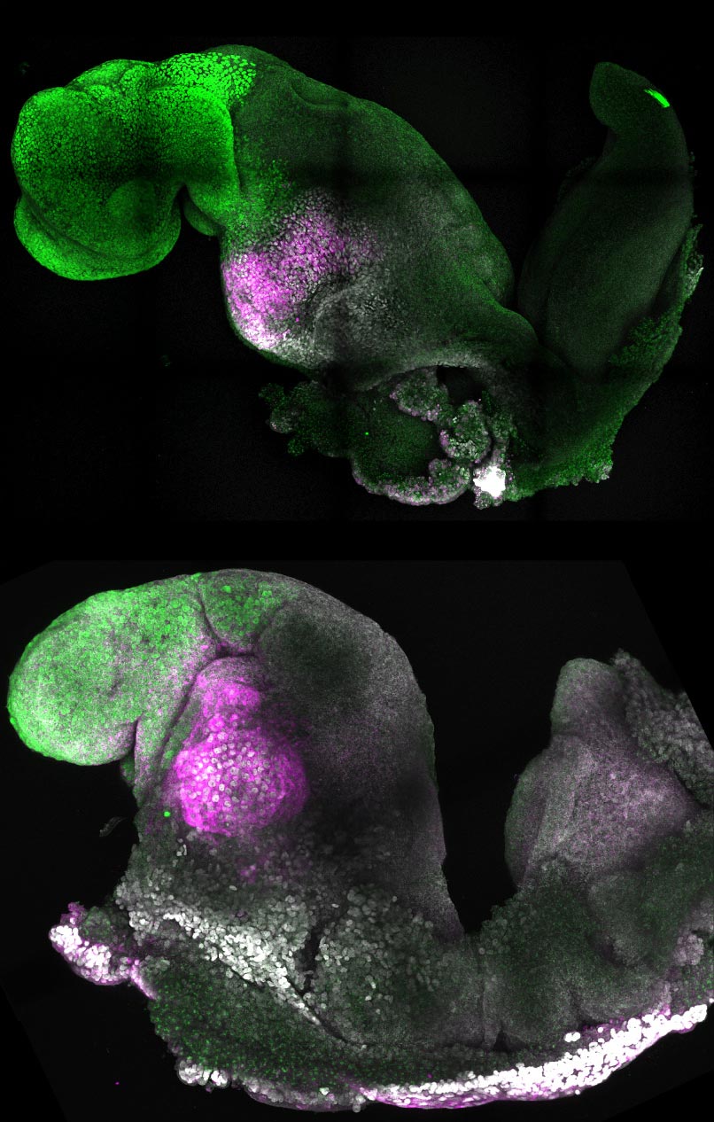
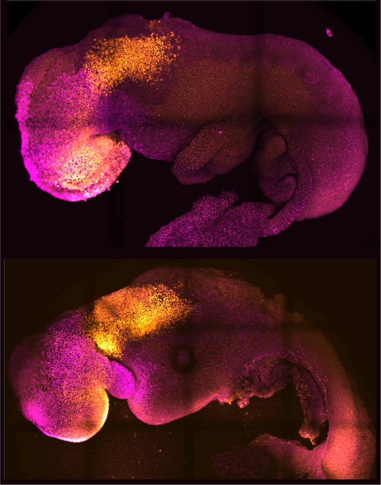
Over time the stem cells self-organized into structures that progressed through the successive developmental stages until the synthetic embryos had beating hearts and the foundations for a brain. They even had the yolk sac where the embryo develops and from which it receives nutrients in its first weeks. This is the most advanced stage of development achieved to date in a stem cell-derived model.
A major advance in this research is the ability to generate the entire brain, in particular the anterior region, which has been a “holy grail” in the development of synthetic embryos.
“This opens new possibilities to study the mechanisms of neurodevelopment in an experimental model,” Zernicka-Goetz says. “In fact, we demonstrate the proof of this principle in the paper by knocking out a gene already known to be essential for formation of the neural tube, precursor of the nervous system, and for brain and eye development. In the absence of this gene, the synthetic embryos show exactly the known defects in brain development as in an animal carrying this mutation. This means we can begin to apply this kind of approach to the many genes with unknown function in brain development.”
“Our mouse embryo model not only develops a brain, but also a beating heart, all the components that go on to make up the body,” she explains. “It’s just unbelievable that we’ve gotten this far. This has been the dream of our community for years, and the major focus of our work for a decade, and finally we’ve done it.”
Studying Early Development and Pregnancy Success Through Synthetic Embryos
For a human embryo to successfully develop, there needs to be a “dialogue” between the tissues that will become the embryo and the tissues that will connect the embryo to the mother. In the first week after fertilization, three types of stem cells develop: one will eventually become the tissues of the body, and the other two will support the embryo’s development. One of these latter two types, known as extraembryonic stem cells, will become the placenta, which connects the fetus to the mother and provides oxygen and nutrients. The other will become the yolk sac, where the embryo grows and from which it receives nutrients in early development.
Many pregnancies fail at the point when the three types of stem cells begin to send mechanical and chemical signals to each other, which tell the embryo how to develop properly.
“This early period is the foundation for everything else that follows in pregnancy,” Zernicka-Goetz says. “If it goes wrong, the pregnancy will fail.”
Over the past decade, Zernicka-Goetz’s team has been investigating these earliest stages of pregnancy to understand why some pregnancies fail and some succeed.
“The stem cell embryo model is important because it gives us accessibility to the developing structure at a stage that is normally hidden from us due to the implantation of the tiny embryo into the mother’s womb,” Zernicka-Goetz says. “This accessibility allows us to manipulate genes to understand their developmental roles in a model experimental system.”
To guide the development of their synthetic embryo, the scientists put together cultured stem cells representing each of the three types of tissue. They allowed them to develop in proportions and an environment conducive to their growth and communication with each other, leading to their eventual self-assembly into an embryo.
The researchers discovered that the extraembryonic cells signal to embryonic cells through chemical signals but also mechanistically, or through touch, guiding the embryo’s development.
“This period of human life is so mysterious, so to be able to see how it happens in a dish—to have access to these individual stem cells, to understand why so many pregnancies fail and how we might be able to prevent that from happening—is quite special,” Zernicka-Goetz says. “We looked at the dialogue that has to happen between the different types of stem cells at that time—we’ve shown how it occurs and how it can go wrong.”
Potential Applications: Synthetic Human Organs and Transplantation
While the current research was carried out in mouse models, the scientists are developing an analogous model for human embryo development to understand mechanisms behind crucial processes that would be otherwise impossible to study in real embryos.
If these methods are demonstrated to be successful with human stem cells in the future, they could also be used to guide the development of synthetic organs for patients awaiting transplants. “There are so many people around the world who wait for years for organ transplants,” Zernicka-Goetz says. “What makes our work so exciting is that the knowledge coming out of it could be used to grow correct synthetic human organs to save lives that are currently lost. It should also be possible to affect and heal adult organs by using the knowledge we have on how they are made.”
Reference: “Synthetic embryos complete gastrulation to neurulation and organogenesis” by Gianluca Amadei, Charlotte E. Handford, Chengxiang Qiu, Joachim De Jonghe, Hannah Greenfeld, Martin Tran, Beth K. Martin, Dong-Yuan Chen, Alejandro Aguilera-Castrejon, Jacob H. Hanna, Michael Elowitz, Florian Hollfelder, Jay Shendure, David M. Glover and Magdalena Zernicka-Goetz, 25 August 2022, Nature.
DOI: 10.1038/s41586-022-05246-3
The paper is titled “Stem cell-derived mouse embryos develop within an extra-embryonic yolk sac to form anterior brain regions and a beating heart.” The co-first authors are Gianluca Amadei and Charlotte Handford of the University of Cambridge. Caltech co-authors are postdoctoral scholars Hannah Greenfeld and Dong-Yuan Chen; graduate student Martin Tran; Michael Elowitz, Professor of Biology and Bioengineering and Howard Hughes Medical Institute Investigator; and David Glover, Research Professor of Biology and Biological Engineering. Additional co-authors are Chengxiang Qiu and Beth Martin of the University of Washington; Joachim De Jonghe and Florian Hollfelder of the University of Cambridge; Alejandro Aguilera-Castrejon and Jacob Hanna of the Weizmann Institute of Science in Israel; and Jay Shendure of the University of Washington, the Brotman Baty Institute for Precision Medicine in Seattle, the Allen Discovery Center for Cell Lineage Tracing in Seattle, and the Howard Hughes Medical Institute in Seattle.
Funding was provided by the National Institutes of Health, the European Research Council, the Wellcome Trust, Open Philanthropy/Silicon Valley Community Foundation and Weston Havens Foundation, and the Centre for Trophoblast Research.
Never miss a breakthrough: Join the SciTechDaily newsletter.
Follow us on Google and Google News.