New research reveals that the cerebral cortex follows the same folding patterns across mammalian species, adhering to a universal fractal shape.
Researchers have developed a novel method for describing the shape of the cerebral cortex, providing evidence that cortices across mammalian species exhibit a universal, fractal pattern.
The study, published as a Reviewed Preprint in eLife and appearing today as a revised version, is described by the editors as a valuable framework to our understanding of the brain cortex as a fractal shape. They describe the strength of evidence as convincing for a universal blueprint for mammalian cerebral cortex folding.
With further research and validation, the approach could be used to grant insights into the development of various degenerative and congenital neuropathic conditions.
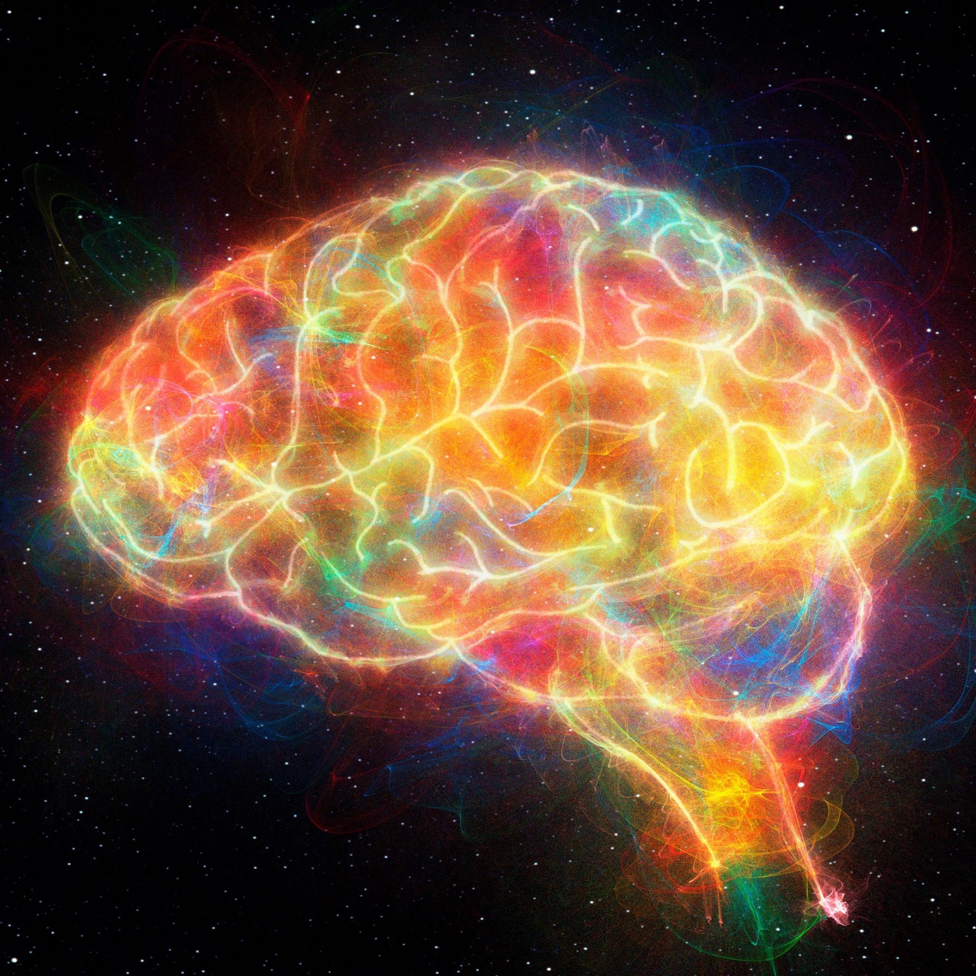
The cerebral cortex is the outermost layer of the brain, and is responsible for complex functions such as thought, perception, and decision-making. Cerebral cortex folding, known as gyrification, is the process by which the brain’s surface develops grooves (sulci) and ridges (gyri). This folding increases the surface area of the brain, allowing for a greater number of neurons and more complex information processing. The cortex displays a wide diversity of shapes and sizes across and within species.
New Methodologies in Cortex Analysis
“We set out to find a way to define the shape of the cortex, and express what is unique about the complex shapes and folds that comprise each cortex,” says lead author Yujiang Wang, a Future Leaders Fellow at the Computational Neurology, Neuroscience & Psychiatry (CNNP) Lab in the School of Computing, Newcastle University, UK. “One can look at an image of a cerebral cortex, and recognize what it is. But how can we tell apart your cortex from mine? Or how can we distinguish a giraffe’s cerebral cortex from that of a marmoset? This requires a more expressive way to describe the shape of the cortex.”
Wang and colleagues began by establishing two key principles. Firstly, they knew that cortices cannot simply assume any folded shape – cortices are thin sheets of grey matter folded in complex ways around white matter, and the degree of folding they undergo is precisely determined by the thickness and size of this sheet. This principle is called universal scaling. They then devised a way to ‘melt’ the cerebral cortex, by removing folds that were smaller than a certain threshold, allowing them to study the remaining folds individually. This revealed the second principle; that cortices are composed of folds of various sizes, where the small folds resemble their larger folds – a property called self-similarity. This resembles fractal scaling, where a complex geometric shape exhibits intricate patterns that repeat at progressively smaller scales.
Comparative Study Across Species
The team then combined these principles of universal scaling and self-similarity to study the cerebral cortex of 11 different primate species, including humans, chimpanzees and marmosets. This revealed that, despite the clear visual differences between the species’ cortices, all of them follow a universal scaling law, and resemble the same fractal shape. So, if you take the most complex cortex studied, that of a human, and use the team’s process of ‘melting’ to eliminate the smallest folds, it begins to resemble that of a chimpanzee. If you ‘melt’ the cortex of a chimpanzee, it resembles that of a rhesus monkey, and so on.
These findings suggest that, regardless of species, there is only one way for a cerebral cortex to undergo folding. So why are they so clearly different when observed through an MRI scan? They appear different in size, and some are highly folded, like the human cortex, and some are much smoother, like the marmoset cortex.
“The key here is to precisely define what we mean by ‘resemble’,” explains senior author Bruno Mota, a Professor at the metaBIO Lab, Instituto de Física, Universidade Federal do Rio de Janeiro, Brazil. “One can imagine a shape that looks like a human cortex, but, as you zoom in, you find within each fold there are infinitely smaller folds. Such a shape cannot exist in nature, but it can be defined mathematically as a fractal shape, as we have done here. What we have shown is that all cortices of the species we have studied resemble this fractal shape for a certain range of fold sizes.”
Therefore, Mota adds, the differences observed in cortical shapes across these species are largely due to the fact that each has a different range of fold sizes for which the resemblance holds. For a smoother cortex, like in a marmoset, this range is narrower; for a more folded one, like a chimpanzee, it is broader.
The authors note that their study was limited to descriptions of entire cortical hemispheres, and that in future work they will look to explore more specific cortical regions. They will also investigate how neurodegenerative diseases such as Alzheimer’s affect the fractal shape of the cortex. This may eventually allow the identification of more detailed biomarkers for various neurological conditions and diseases, and grant further understanding for how they develop.
“Our results suggest a universal blueprint for mammalian brain shape, and a common set of mechanisms governing cortical folding,” concludes Mota. “We hope that our framework for expressing and analyzing cortical shape can become a powerful tool to characterize and compare cortices of different species and individuals, across development and aging, and across health and disease.”
Reference: “Neuro-evolutionary evidence for a universal fractal primate brain shape” by Yujiang Wang, Karoline Leiberg, Nathan Kindred, Christopher R. Madan, Colline Poirier, Christopher I. Petkov, Peter N. Taylor and Bruno Mota, 30 July 2024, eLife.
DOI: 10.7554/eLife.92080.3
Never miss a breakthrough: Join the SciTechDaily newsletter.
Follow us on Google and Google News.
1 Comment
That was about what I would expect as far as human behaviors are concerned, “blame it on the folds or some other such thing”; well, that’s great, next time we pay more attention to the folds; this species obviously, can’t past inspection because we don’t have the technology yet to perform an adequate quality control inspection. So then; what? We just toss all these 8 billion people into the recycling bin because they don’t pass inspection?
Just a joke, but obviously on some scale we’re all unusable junk.