

AI-based tissue clocks reveal that human organs age on different timelines and that some of these patterns can be detected from blood.
The age printed on a birth certificate tells only part of the story. Inside the body, the lungs, kidneys, pancreas, brain, and other organs may be aging along very different timelines, and artificial intelligence can now detect some of those differences in the microscopic structure of human tissue.
Researchers at the CeMM Research Center for Molecular Medicine of the Austrian Academy of Sciences and the Ludwig Boltzmann Institute for Network Medicine (LBI-NetMed) at the University of Vienna developed AI-based “tissue clocks” that estimate the biological age of individual organs from histology images.
By examining more than 25,000 images spanning 40 tissue types, they found that organs do not age uniformly and that some of these aging patterns can also be inferred from blood. The findings were published in Nature Medicine and could eventually contribute to disease monitoring and earlier diagnosis.
The familiar observation that two people of the same chronological age can appear to age differently raises a question: are their individual organs also aging at different rates? And can researchers measure the gap between a person’s chronological age and the biological age of a particular tissue?
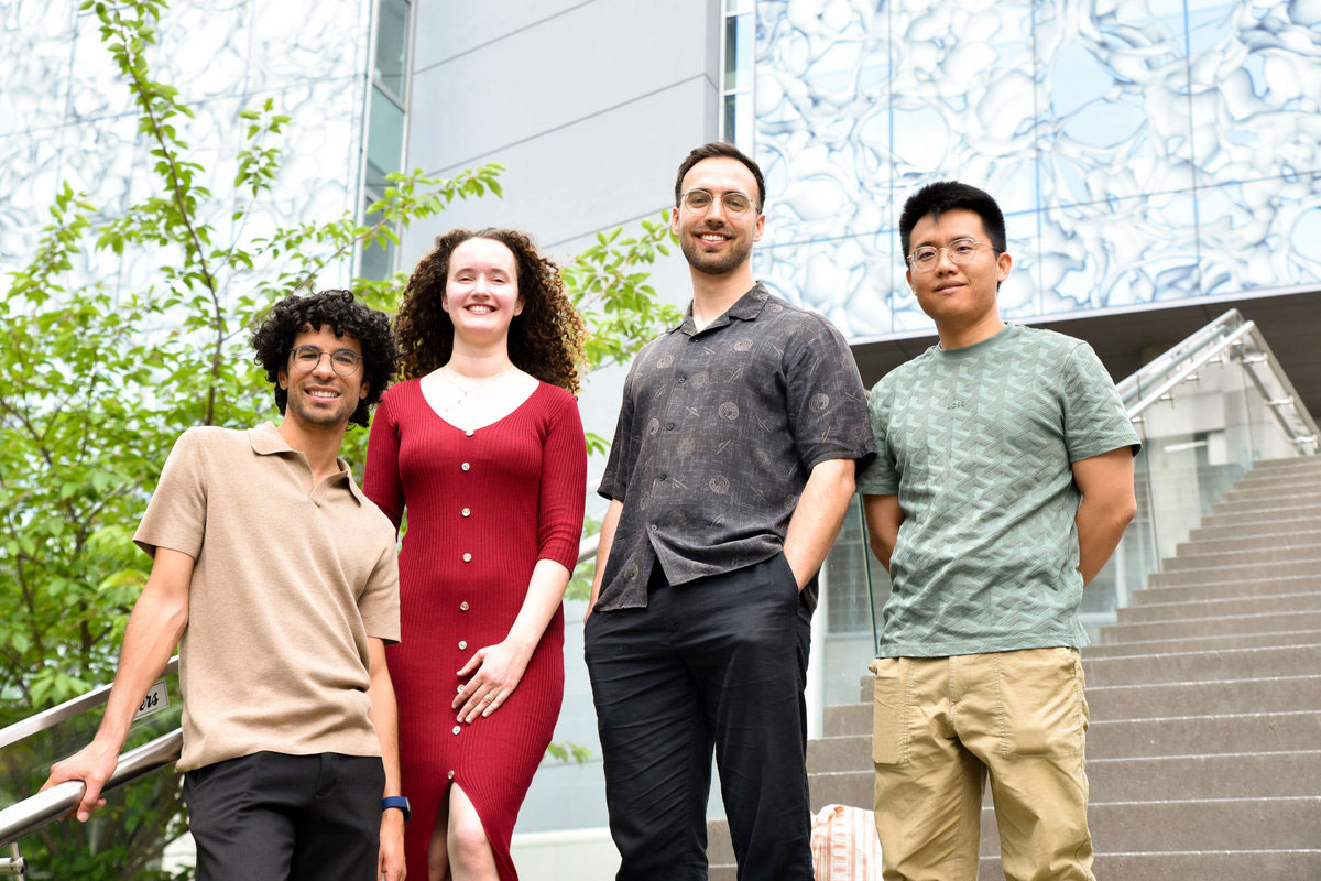
To investigate, CeMM and LBI-NetMed Principal Investigator André Rendeiro and co-first authors Ernesto Abila, Iva Buljan, and Yimin Zheng combined artificial intelligence with one of the world’s largest collections of human tissue images. Earlier approaches to biological aging have often focused on molecular changes such as DNA methylation or gene expression. This study instead asked whether the physical architecture of tissues contains its own record of aging.
AI reads age from tissue architecture
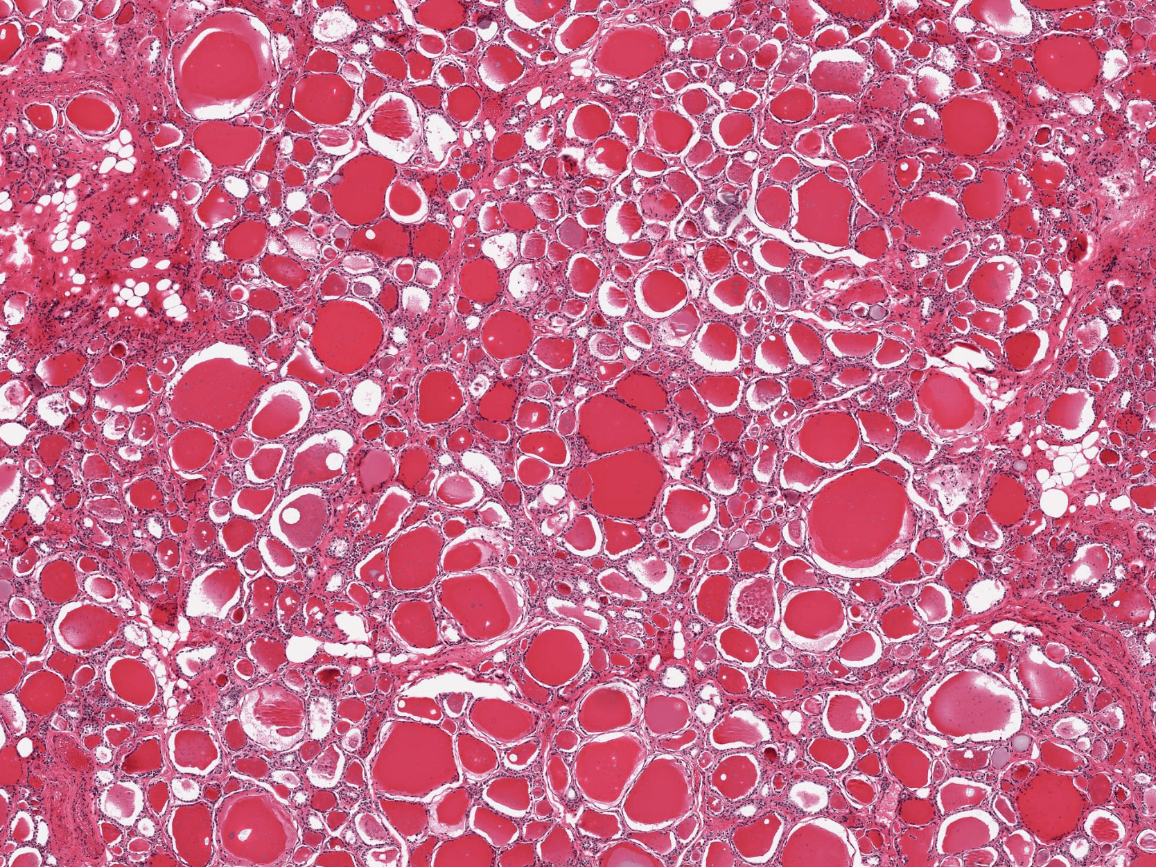
The researchers used data from the Genotype-Tissue Expression Project (GTEx), which collected samples from 983 people across 40 tissue types, including the brain, heart, lung, pancreas, skin, and intestine. Those samples were converted into high-resolution digital images of tissue sections showing the microscopic organization of each organ. In total, the researchers analyzed 25,712 images representing about 480 million individual image tiles with advanced computer vision models.
Even though the models had not been explicitly trained to look for aging, age proved to be the strongest factor influencing tissue appearance across all 40 tissue types. The researchers then used those patterns to construct ’tissue clocks,’ predictive models capable of estimating biological age separately for each organ based on its microscopic appearance.
Across tissues, the clocks had an average prediction error of 4.9 years. They also outperformed existing DNA-based aging estimates at capturing tissue-specific pathology. The estimated biological ages were strongly associated with established features of aging, including shorter telomeres, tissue pathology, and the number of chronic diseases a person had.
“Our tissues carry a remarkably detailed record of the aging process,” says André Rendeiro, Principal Investigator at CeMM and corresponding author of the study. “By combining histology images with artificial intelligence, we can detect patterns of biological aging that are invisible to the human eye and begin to understand how aging unfolds differently across the body.”
Organs age on different timelines
The results showed that aging follows different trajectories depending on the tissue. The lung, kidney, pancreas, and adrenal gland displayed signs of accelerated aging as early as ages 20 to 40, while other tissues showed more complex patterns with later peaks. The uterus underwent a particularly pronounced shift around menopause.
The researchers also found connections between tissue-specific aging and medical conditions or lifestyle-associated factors. Kidney failure, for example, was associated with accelerated aging signals across several tissues, while diabetes showed especially strong effects in the pancreas.
“What stands out is how differently each organ ages, and how that shows up in tissue architecture,” says Ernesto Abila, co-first author of the study. “Deep learning lets us read these spatial patterns, capturing aging as architectural remodeling, not just molecular drift.” The tissue clocks captured typical aging patterns across organs while also identifying individuals whose tissues showed pronounced structural changes earlier than expected for their chronological age.
Because collecting tissue is not always practical, the researchers next asked whether those organ-specific aging patterns could be detected in blood. They matched blood-based gene expression profiles with tissue age gaps measured from histology in the same individuals and used the combined information to build predictors of tissue-specific biological age from blood alone.
“This is a conceptual leap: using the language of tissue aging, learned from images, and translating it into something readable from a routine blood draw,” explains co-first author Iva Buljan.
The blood-based predictors identified aging patterns associated with Alzheimer’s disease, Crohn’s disease, cystic fibrosis, vasculitis, diabetes, and stroke. In people with Alzheimer’s disease, the strongest aging signal appeared in the brain, while Crohn’s disease was associated with accelerated aging across the gastrointestinal tract.
Tissue aging could inform future diagnostics
“This study highlights that aging is not simply a matter of chronological time,” says Yimin Zheng, the third co-first author of the study. “Different organs age in different ways, and these processes appear to be shaped by both systemic and tissue-specific factors.” The findings suggest that tissue architecture reflects many of the molecular and physiological changes associated with both aging and disease. In the future, similar approaches could contribute to minimally invasive tests that track organ health and disease progression through blood samples.
The work also illustrates how artificial intelligence can connect pathology, gene expression, and clinical information at large scale. By combining these different layers of data, the researchers produced a more detailed view of how aging varies across the human body.
Reference: “Histological aging signatures for monitoring tissue-specific aging and disease” by Ernesto Abila, Iva Buljan, Yimin Zheng (郑易民), Lisa Kleissl, Sigrid Klotz, Tamas Veres, Zhilong Weng, Maja Nackenhorst, Rizqah Kamies, Anja Michl, Safwen Kadri, Samir Moustafa, Wolfgang Hulla, Matthias Perkonigg, Mathias Drach, Philipp Tschandl, Barbara Sterniczky, Matthias Heinig, Laurens J. De Sadeleer, Wim Wuyts, Bart Vanaudenaerde, Laurens J. Ceulemans, Daniel D. Buchanan, Lochlan J. Fennell, Georg Stary, Yuri Tolkach, Adelheid Wöhrer, Herbert B. Schiller and André F. Rendeiro, 14 August 2026, Nature Medicine.
DOI: 10.1038/s41591-026-04566-5
The GTEx project was supported by the Common Fund of the Office of the Director of the National Institutes of Health and by the National Cancer Institute; National Human Genome Research Institute; National Heart, Lung and Blood Institute; National Institute on Drug Abuse; National Institute of Mental Health and National Institute of Neurological Disorders and Stroke.
Never miss a breakthrough: Join the SciTechDaily newsletter.
Follow us on Google and Google News.