Proteins in the sheath of cellular protrusions control how effectively cells can adhere to surfaces.
Biological cells often feature thin, hair-like structures on their surface called cilia, which play key roles in movement and sensing environmental signals. A team of researchers from Germany and Italy has recently uncovered new details about the protective layer that surrounds these cilia.
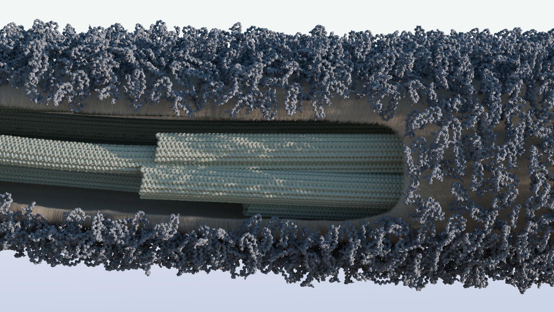
This layer, known as the glycocalyx, is composed of sugar-rich proteins called glycoproteins. As the cell’s first point of contact with its environment, the glycocalyx influences how cells adhere to surfaces, move, and detect external signals. Until now, its precise structure remained unclear. The research team has now successfully mapped the detailed architecture of the glycocalyx in the unicellular green alga Chlamydomonas reinhardtii, identifying the glycoproteins FMG1B and FMG1A as its primary components.
FMG1A is a previously unknown variant of FMG1B, and the two glycoproteins show a biochemical similarity to mucin proteins found in mammals. Mucins are also glycoproteins and a central component of protective mucus found in many organisms, for example, on mucous membranes or in internal organs.
Functional Role of the Glycoproteins
For their study, the team removed the two glycoproteins from the alga, which resulted in the cilia showing significantly increased stickiness. Nonetheless, the algal cells were still able to move on surfaces by means of the adhering cilia. This led the researchers to conclude that these proteins do not, as previously assumed, directly enable adhesion to surfaces and transmit the force needed for gliding motility from inside the cilium, but instead form a protective layer that regulates the adhesiveness of the cilia.
“This discovery expands our knowledge of how cells regulate direct interaction with their environment,” explains plant biotechnologist Prof Michael Hippler from the University of Münster (Germany).
“We are also gaining insights into how similar protective mechanisms might work in other organisms,” adds Dr Adrian Nievergelt from the Max Planck Institute of Molecular Plant Physiology in Potsdam (Germany), who collaborated on the project with Dr Gaia Pigino’s research group at the Human Technopole in Milan (Italy).
The team used a wide range of cutting-edge imaging and protein analysis techniques, including cryogenic electron tomography and electron microscopy, fluorescence microscopy, mass spectrometry, as well as genetic manipulation to remove the glycoproteins from the algal genome.
Reference: “Unwrapping the Ciliary Coat: High-Resolution Structure and Function of the Ciliary Glycocalyx” by Lara M. Hoepfner, Adrian P. Nievergelt, Fabrizio Matrino, Martin Scholz, Helen E. Foster, Jonathan Rodenfels, Alexander von Appen, Michael Hippler and Gaia Pigino, 5 March 2025, Advanced Science.
DOI: 10.1002/advs.202413355
Funding: European Research Council, Deutsche Forschungsgemeinschaft, Human Frontier Science Program, European Molecular Biology Organization
Never miss a breakthrough: Join the SciTechDaily newsletter.
Follow us on Google and Google News.