Zebrafish Reveal Potential for Heart Regeneration in Humans
Cardiovascular conditions like heart attacks are among the major causes of death globally, a result of the human heart’s limited self-healing power. Unlike humans, zebrafish possess a remarkable ability to recover from heart injuries.
A team of researchers led by Jeroen Bakkers from the Hubrecht Institute has utilized zebrafish to uncover the secrets behind their regenerative abilities. They identified a novel mechanism that acts as a trigger, prompting the maturation of heart muscle cells during the regeneration process. Importantly, this mechanism was evolutionarily conserved as it had a very similar effect on mouse and human heart muscle cells.
The results of the study, published in Science on May 18th, show that examining the natural heart regeneration process in zebrafish and applying these discoveries to human heart muscle cells could contribute to the development of new therapies against cardiovascular diseases.
It is estimated that 18 million people die from cardiovascular diseases every year. Many of these deaths are related to heart attacks. In such an event, a blood clot prevents the supply of nutrients and oxygen to parts of the heart. As a result, the heart muscle cells in the obstructed part of the heart die, which eventually leads to heart failure.
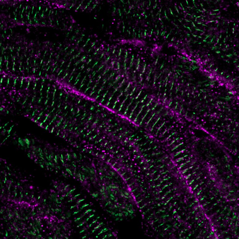
Live imaging of calcium movements through zebrafish heart muscle cells 21 days after injury. Credit: Phong Nguyen, copyright: Hubrecht Institute.
Although therapies exist that manage the symptoms, there is no treatment that is able to replace the lost tissue with functional, mature heart muscle cells and thereby cure the patients.
Zebrafish as a Role Model
Unlike humans, some species like zebrafish can regenerate their hearts. Within 90 days after damage, they fully restore their cardiac function. The surviving heart muscle cells are able to divide and produce more cells. This unique feature provides zebrafish hearts with a source of new tissue to replace the lost heart muscle cells. Previous studies successfully identified factors that could stimulate heart muscle cells to divide. Nevertheless, what happens to the newly formed heart muscle cells afterward had not been studied before.
Phong Nguyen, first author of the study, explains: “It is unclear how these cells stop dividing and mature enough so that can they contribute to normal heart function. We were puzzled by the fact that in zebrafish hearts, the newly formed tissue naturally matured and integrated into the existing heart tissue without any problems.”
LRRC10 Drives Maturation
To study the maturation of the newly formed tissue in detail, the researchers developed a technique for which thick slices of injured zebrafish hearts were cultured outside the body. This allowed them to perform live imaging on the movement of calcium in heart muscle cells.
The regulation of calcium moving in and out of heart muscle cells is important for controlling heart contractions and can predict the maturity of the cell. They found that after the heart muscle cells divide, calcium movements changed over time.
Live imaging of calcium movements through lab-grown human heart muscle cells (hiPSC-CM). Credit: Phong Nguyen and Giulia Campostrini, copyright: Hubrecht Institute.
“The calcium movement in the newly divided cell was initially very similar to embryonic heart muscle cells, but over time the heart muscle cells assumed a mature type of calcium movement. We found that the cardiac dyad, a structure that helped to move calcium within the heart muscle cell, and specifically one of its components, LRRC10, was crucial in deciding whether heart muscle cells divide or progress through maturation. Heart muscle cells that lack LRRC10 continued to divide and remained immature,” says Nguyen.
From Fish to Human
After Nguyen and his colleagues established the importance of LRRC10 in stopping cell division and initiating the maturation of zebrafish heart muscle cells, they moved on to test if their findings could be translated to mammals. To this end, they induced the expression of LRRC10 in mouse and lab-grown human heart muscle cells.
Strikingly, LRRC10 changed the calcium handling, reduced cell division, and increased the maturation of these cells in a similar manner as observed in zebrafish hearts.
Nguyen: “It was exciting to see that the lessons learned from the zebrafish were translatable as this opens new possibilities for the use of LRRC10 in the context of new therapies for patients.”
Clinical Impact
The results of the study, published in Science, show that LRRC10 has the potential to drive the maturation of heart muscle cells further through the control of their calcium handling. This could help scientists who are trying to solve the lack of regenerative capacity of the mammalian heart by transplanting lab-grown heart muscle cells into the damaged heart.
Although this potential therapy is promising, results showed that these lab-grown cells are still immature and cannot communicate properly with the rest of the heart, leading to abnormal contractions called arrhythmias.
“Although more research is needed to precisely define how mature these lab-grown heart muscle cells are when treated with LRRC10, it is possible that the increase in maturation will improve their integration after transplantation,” says Jeroen Bakkers, last author of the study.
Bakkers continues: “Additionally, current models for cardiac diseases are frequently based on immature lab-grown heart muscle cells. 90% of promising drug candidates found in the lab fail to make it to the clinic and the immaturity of these cells could be one contributing factor for this low success rate. Our results indicate LRRC10 could improve the relevance of these models as well.”
LRRC10 could thus have an important contribution to generating lab-grown heart muscle cells that more accurately represent a typical adult human heart, therefore improving the chances of developing successful new treatments against cardiovascular diseases.
Reference: “Interplay between calcium and sarcomeres directs cardiomyocyte maturation during regeneration” by Phong D. Nguyen, Iris Gooijers, Giulia Campostrini, Arie O. Verkerk, Hessel Honkoop, Mara Bouwman, Dennis E. M. de Bakker, Tim Koopmans, Aryan Vink, Gerda E. M. Lamers, Avraham Shakked, Jonas Mars, Aat A. Mulder, Sonja Chocron, Kerstin Bartscherer, Eldad Tzahor, Christine L. Mummery, Teun P. de Boer, Milena Bellin and Jeroen Bakkers, 18 May 2023, Science.
DOI: 10.1126/science.abo6718
The study is the result of a collaboration between the Hubrecht Institute, LUMC, AMC, UMC Utrecht and Weizmann Institute. The study was financed with the help of the Dutch Heart Foundation, Dutch CardioVascular Alliance, and Stichting Hartekind.
Funding: European Molecular Biology Organization, Human Frontier Science Program, NWO-ZonMW Veni grant, Horizon 2020 Framework Programme, Netherlands Organ-on-Chip Initiative, an NWO Gravitation project funded by the Ministry of Education, Culture and Science of the government of the Netherlands, European Research Council, Novo Nordisk Foundation Center for Stem Cell Medicine supported by Novo Nordisk Foundation grants, European Research Council, Netherlands Cardiovascular Research Initiative: An initiative with support of the Dutch Heart Foundation and Hartekind, NWO-ZonMW Open competition grant CONTRACT
Never miss a breakthrough: Join the SciTechDaily newsletter.
Follow us on Google and Google News.
1 Comment
……… AND, how do you repair the damaged heart. Did not say.