A recent study has discovered a mechanism for controlling calcium influx in cells that can help researchers understand the molecular reasons behind the disruption of brain functioning in neurological disorders, including stroke.
In a newly published study, scientists identify the mechanism for controlling calcium influx in cells, helping researchers better understand the molecular causes of the disruption of brain functioning that occurs in stroke and other neurological disorders.
When brain cells are overwhelmed by an influx of too many calcium molecules, they shut down the channels through which these molecules enter the cells. Until now, the “stop” signal mechanism that cells use to control the molecular traffic was unknown.
In the new issue of the journal Neuron, UC Davis Health System scientists report that they have identified the mechanism. Their findings are relevant to understanding the molecular causes of the disruption of brain functioning that occurs in stroke and other neurological disorders.
“Too much calcium influx clearly is part of the neuronal dysfunction in Alzheimer’s disease and causes the neuronal damage during and after a stroke. It also contributes to chronic pain,” said Johannes W. Hell, professor of pharmacology at UC Davis. Hell headed the research team that identified the mechanism that stops the flow of calcium molecules, which are also called ions, into the specialized brain cells known as neurons.
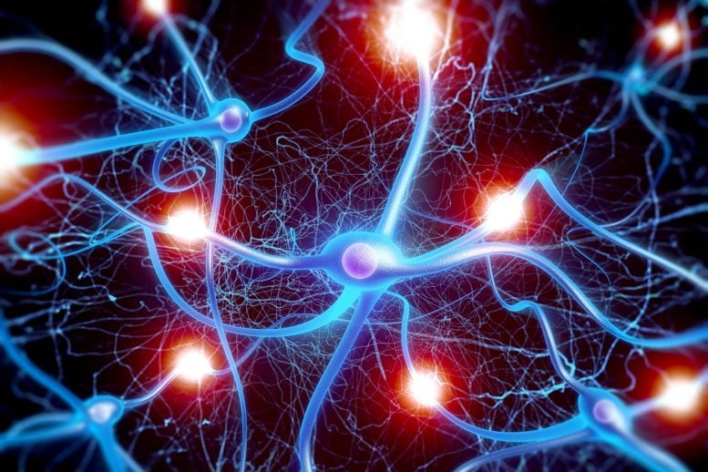
Hell explained that each day millions of molecules of calcium enter and exit each of the 100 billion neurons of the human brain. These calcium ions move in and out of neurons through pore-like structures, known as channels, that are located in the outer surface, or “skin,” of each cell.
The flow of calcium ions into brain cells generates the electrical impulses needed to stimulate such actions as the movement of muscles in our legs and the creation of new memories in the brain. The movement of calcium ions also plays a role in gene expression and affects the flexibility of the structures, called synapses, that are located between neurons and transmit electrical or chemical signals of various strengths from one cell to a second cell.
Neurons employ an unexpected and highly complex mechanism to down regulate, or reduce, the activity of channels that are permitting too many calcium ions to enter neurons, Hell and his colleagues discovered. The mechanism, which leads to the elimination of the overly permissive ion channel employs two proteins, α-actinin and the calcium-binding messenger protein calmodulin.
Located on the neuron’s outer surface, referred to as the plasma membrane, α-actinin stabilizes the type of ion channels that constitute a major source of calcium ion influx into brain cells, Hell explained. This protein is a component of the cytoskeleton, the scaffolding of cells. The ion channels that are a major source of calcium ions are referred to as Cav1.2 (L type voltage-dependent calcium channels).
The researchers also found that the calcium-binding messenger protein calmodulin, which is the cell’s main sensor for calcium ions, induces internalization, or endocytosis, of Cav1.2 to remove this channel from the cell surface, thus providing an important negative feedback mechanism for excessive calcium ion influx into a neuron, Hell explained.
The discovery that α-actinin and calmodulin play a role in controlling calcium ion influx expands upon Hell’s previous research on the molecular mechanisms that regulate the activity of various ion channels at the synapse.
One previous study proved relevant to understanding the biological mechanisms that underlie the body’s fight-or-flight response during stress.
In work published in the journal Science in 2001, Hell and colleagues reported that the regulation of Cav1.2 by adrenergic signaling during stress is performed by one of the adrenergic receptors (beta 2 adrenergic receptor) directly linked to Cav1.2.
“This protein-protein interaction ensures that the adrenergic regulation is fast, efficient and precisely targets this channel,” Hell said.
“We showed that Cav1.2 is regulated by adrenergic signaling on a time scale of a few seconds, and this is mainly increasing its activity when needed, for example during danger, to make our brain work faster and better. The same channel is in the heart, where adrenergic stimulation increases channel/Ca influx activity, increasing the pacing and strength of our heartbeat to meet the increased physical demands during danger.”
Reference: “Competition between α-actinin and Ca2+-Calmodulin Controls Surface Retention of the L-type Ca2+ Channel CaV1.2” by Duane D. Hall, Shuiping Dai, Pang-Yen Tseng, Zulfiqar Malik, Minh Nguyen, Lucas Matt, Katrin Schnizler, Andrew Shephard, Durga P. Mohapatra, Fuminori Tsuruta, Ricardo E. Dolmetsch, Carl J. Christel, Amy Lee, Alain Burette, Richard J. Weinberg and Johannes W. Hell, 8 May 2013, Neuron.
DOI: 10.1016/j.neuron.2013.02.032
Other authors on the paper include Duane D. Hall, Shuiping Dai, Katrin Schnizler, Andrew Shephard, Durga P. Mohapatra, Carl J. Christel and Amy Lee from the University of Iowa; Pang-Yen Tseng, Zulfiqar Malik, Minh Nguyen, Lucas M. Matt from UC Davis; Fuminori Tsuruta and Ricardo E. Dolmetsch from Stanford University; and Alain Burette and Richard Weinberg from the University of North Carolina.
This work was supported by National Institutes of Health training grants HL07121, DK07759, AG00213, AG017502, NS35527, DC009433 and HL087120 to AL, as well as the American Heart Association grant 0535235N.


Be the first to comment on "Mechanism for Controlling Calcium Influx in Cells Identified"