
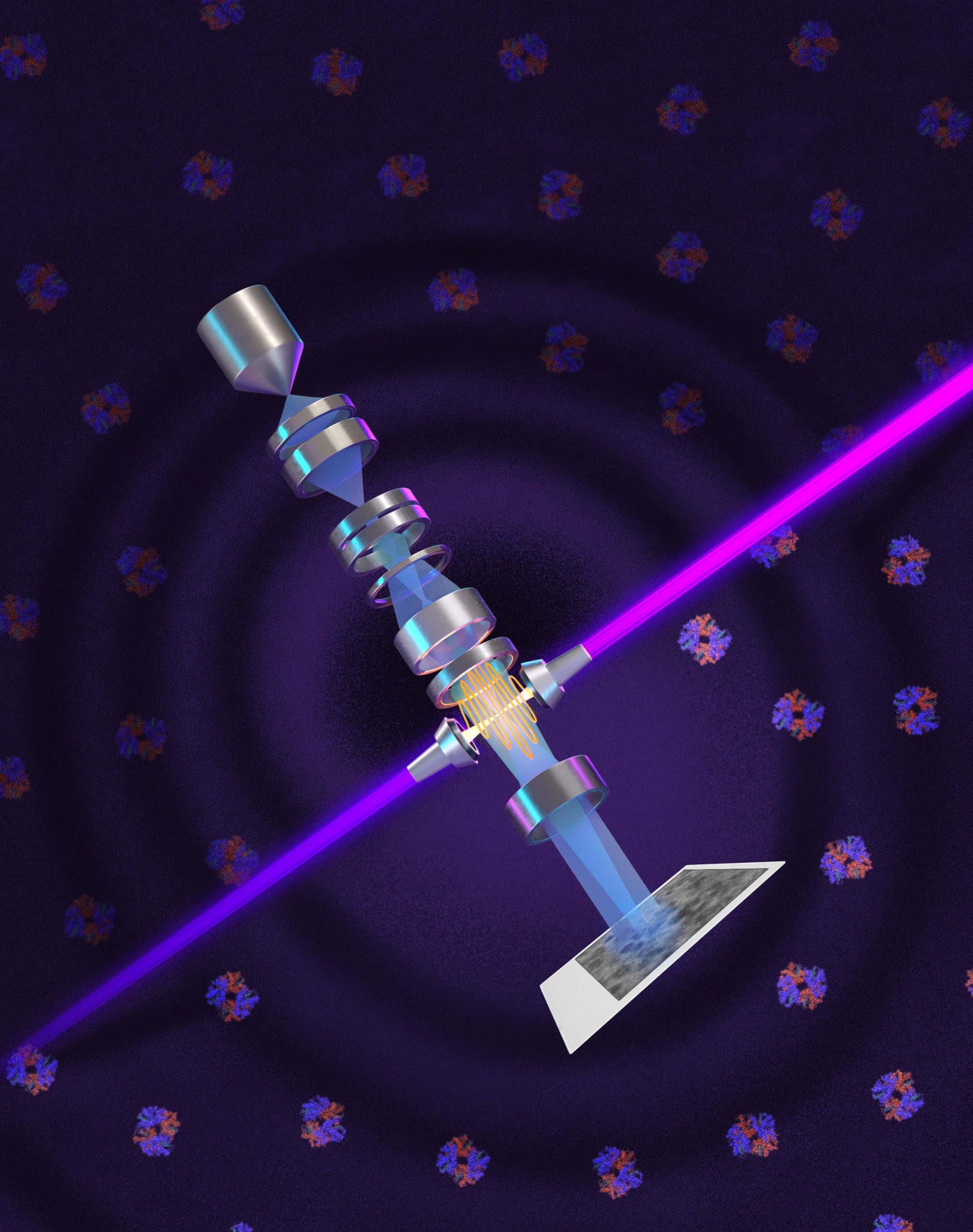
A laser-based innovation could allow electron microscopes to reveal biological structures that have long remained beyond their reach.
Most human proteins remain too small for today’s most powerful biological microscopes to resolve clearly. A laser system developed by UC Berkeley physicists could help bring many of them into view.
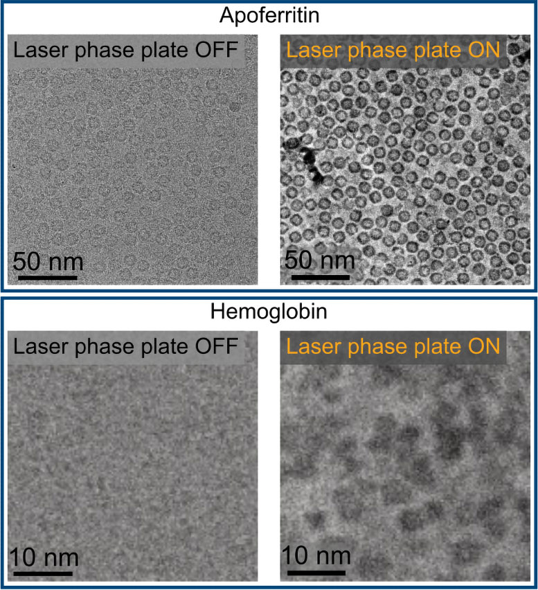
The researchers adapted phase contrast, a nearly century-old imaging method, for cryoelectron microscopy (cryo-EM). Their laser phase plate improves contrast by changing the phase of an electron beam without significantly reducing its intensity.
The advance could also strengthen cryoelectron tomography (cryo-ET), which combines images taken from different angles to reconstruct molecules inside cells.
Revealing Smaller Proteins
“Cryo-EM has become the new, fastest-growing method for resolving the structure of biological macromolecules, and cryo-ET is expected to show how these molecules work together in their natural, cellular context,” said Holger Müller, a UC Berkeley professor of physics and faculty scientist at Lawrence Berkeley National Laboratory who led the development effort. “But because of signal-to-noise limitations, the majority of human and animal proteins are too small to be analyzed by these methods. The increase in signal-to-noise ratio provided by this laser phase plate is expected to overcome these important limitations.”
The system uses an exceptionally intense focused continuous-wave laser to alter the electron beam. This change makes small molecules such as hemoglobin easier to distinguish and could improve views of crowded structures inside cells.
“With cryo-ET, we’re looking at small, very complicated cellular material that’s incredibly crowded inside the cell,” said Bridget Carragher, founding technical director of imaging at Biohub in Redwood City, California. “It’s like a forest of trees, and you’re trying to find one leaf on one tree in there. Cryo-ET needs a dramatic step forward in contrast, so we can start to see what’s going on inside the cell. That’s what the laser phase plate promises to give us.”
Building Theia
Biohub funded a customized Thermo Fisher Krios microscope that Müller equipped with the laser phase plate. He named the instrument Theia after the ancient Greek Titaness associated with light.
Biohub is developing a second microscope that uses two perpendicular lasers at lower power. The design is intended to reduce optical distortions and protect components from damage.
“Theia is the Formula 1 microscope,” Müller said. “It has extra electron optics that give it better resolution than the standard cryo-EM, even without the laser. With the addition of the laser phase plate, we hope that it really becomes the world’s best instrument overall.”
A Century-Old Idea Reworked
Phase contrast was developed by Dutch physicist Frits Zernike in 1930. He discovered that light passing through biological material changes not only in brightness but also in phase.
By shifting unscattered light by 90 degrees, phase contrast converts nearly invisible differences into changes in brightness. The method made transparent cellular structures easier to see without staining them. Zernike received the 1953 Nobel Prize in Physics for the discovery.
Scientists soon tried to apply the same principle to electron microscopes, but early phase plates weakened the beam, reduced resolution, or produced unstable images.
In 2010, Müller and cryo-EM pioneer Robert Glaeser proposed using an intense laser instead.
Fifteen Years of Development
Müller spent 15 years turning the concept into a working instrument. His team trapped a laser inside a spherical mirrored cavity, where the light reflects more than 10,000 times and becomes concentrated into a tiny area.
“It’s 75 kilowatts focused to a few microns,” Müller said. “That’s more powerful than what you use for welding. It’s more power than a military laser. It builds up the brightest continuous laser focus ever.”
Tests involving six biological samples showed that the laser produced the greatest improvements for small or difficult specimens.
“For the most challenging cases — small particles, bad specimens — the laser produces a very considerable advantage,” Müller said.
The researchers tested the system on aldolase, a muscle protein that cryo-EM can already image relatively well, and hemoglobin, a smaller protein near the lower limit of current instruments. Both images improved, but hemoglobin benefited more.
Pushing Beyond Current Limits
Cryo-EM can barely resolve proteins smaller than about 70 kilodaltons, even though proteins below that size account for roughly 90 percent of the human proteome.
With the laser phase plate, researchers can now image proteins as small as 50 kilodaltons, although doing so remains difficult. Müller hopes future improvements will lower the limit to 17 kilodaltons (the size of the protein myoglobin).
A focused electron beam could provide an additional twofold improvement in contrast and signal-to-noise ratio.
“This technology is a step function change for biology,” said Stephani Otte, Biohub’s Vice President of Imaging Science. “We are going to be able to see how molecular machines operate inside the living cell, in context, for the first time. What was once invisible will become visible — and that changes everything about how we understand disease.”
Reference: “Laser phase plate improves structure determination of small proteins by cryo-EM” by Petar N. Petrov, Jessie T. Zhang, Jonathan Remis, Jeremy J. Axelrod, Hang Cheng, Eric S. Cooper, Ian K. Hicklin, Shahar Sandhaus, Cooper Schnurr, Robert M. Glaeser and Holger Müller, 11 June 2026, Science.
DOI: 10.1126/science.aeh0665
Never miss a breakthrough: Join the SciTechDaily newsletter.
Follow us on Google and Google News.
3 Comments
“The laser phase plate finally treats the electron beam like a high-precision industrial inspection tool. A flat 2D shadow always implies a volumetric source, but for decades, standard microscopy crushed that depth information, forcing academics to rely on computer algorithms to ‘guess’ protein shapes. By using a localized energy gradient to amplify and square up the wave phase, this breakthrough restores the missing Z-axis. It proves that when our tools have the proper mechanical leverage, the theoretical mysteries disappear, leaving nothing but undeniable 3D geometry on the workbench.”
What is the difference between death and living 3D? Is there absolute stillness?
A laser-based innovation could allow electron microscopes to reveal biological structures that have long remained beyond their reach. We are going to be able to see how molecular machines operate inside the living cell, for the first time.
VERY GOOD!
Please ask researchers to think deeply:
What is the difference in biological structure between living cells and dead cells?