Close Menu
    Facebook X (Twitter) Instagram
    SciTechDaily
    • Biology
    • Chemistry
    • Earth
    • Health
    • Physics
    • Science
    • Space
    • Technology
    Facebook X (Twitter) Pinterest YouTube RSS
    SciTechDaily
    Home»Health»MRI Reveals Significantly Altered Brain Structure in Fetuses Exposed to Alcohol
    Health

    MRI Reveals Significantly Altered Brain Structure in Fetuses Exposed to Alcohol

    By Radiological Society of North AmericaDecember 1, 2021No Comments5 Mins Read
    Facebook Twitter Pinterest Telegram LinkedIn WhatsApp Email Reddit
    Share
    Facebook Twitter LinkedIn Pinterest Telegram Email Reddit
    MRI Reveals Altered Brain Structure in Fetuses Exposed to Alcohol
    MRI super-resolution reconstruction and atlas-based tissue segmentation. (A, B) Postprocessed MRI super-resolution reconstructions in axial and sagittal planes of a fetus at 26+6 GW. (C, D) Respective manually corrected atlas-based tissue segmentation. Color coding: blue — external CSF-spaces, red — cortex, orange — subcortical parenchyma, brown — periventricular zone, dark green — ganglionic eminence, white — ventricular system, dark blue — corpus callosum, yellow – deep gray nuclei (basal ganglia and thalamus), light green — brainstem, light blue — cerebellum, turquoise — left hippocampus, gray — right hippocampus. Credit: RSNA and Marlene Stuempflen, M.D.

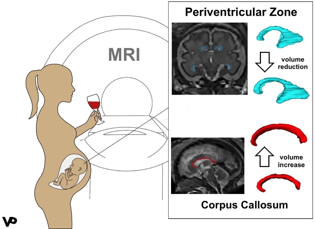
    MRI scans reveal that prenatal alcohol exposure alters fetal brain structures, including enlargement of the corpus callosum and reduced volume in the periventricular zone.

    In the first MRI-based study to investigate prenatal alcohol exposure, researchers found significant changes in the brain structure of fetuses exposed to alcohol compared to healthy controls. Results of the study are being presented today at the annual meeting of the Radiological Society of North America (RSNA).

    “Fetal alcohol syndrome is a worldwide problem in countries where alcohol is freely available,” said Gregor Kasprian, M.D., associate professor of radiology at the Medical University of Vienna in Austria. “It’s estimated that 9.8% of all pregnant women are consuming alcohol during pregnancy, and that number is likely underestimated.”

    Fetal alcohol syndrome is the most severe form of a group of conditions called fetal alcohol spectrum disorders that result from alcohol exposure during pregnancy. Babies born with fetal alcohol spectrum disorders may have specific physical features, learning disabilities, behavioral problems or speech and language delays. According to Dr. Kasprian, one in 70 pregnancies with alcohol exposure results in fetal alcohol syndrome.

    “There are many postnatal studies on infants exposed to alcohol,” Dr. Kasprian said. “We wanted to see how early it’s possible to find changes in the fetal brain as a result of alcohol exposure.”

    Effects of Prenatal Alcohol Exposure
    Graphical abstract: effects of prenatal alcohol exposure (PAE). PAE leads to a volume reduction in the periventricular zone and a volume increase in the corpus callosum. Credit: RSNA and Marlene Stuempflen, M.D.

    For the study, researchers recruited 500 pregnant women who were referred for a fetal MRI for clinical reasons. On an anonymous questionnaire, 51of the women admitted to consuming alcohol during their pregnancy. The questionnaires used were the Pregnancy Risk Assessment Monitoring System (PRAMS), a surveillance project of the Centers for Disease Control and Prevention and health departments, and the T-ACE Screening Tool, a measurement tool of four questions that identify risk drinking.

    “We provided a safe environment where women could feel comfortable honestly answering the questions,” Dr. Kasprian said.

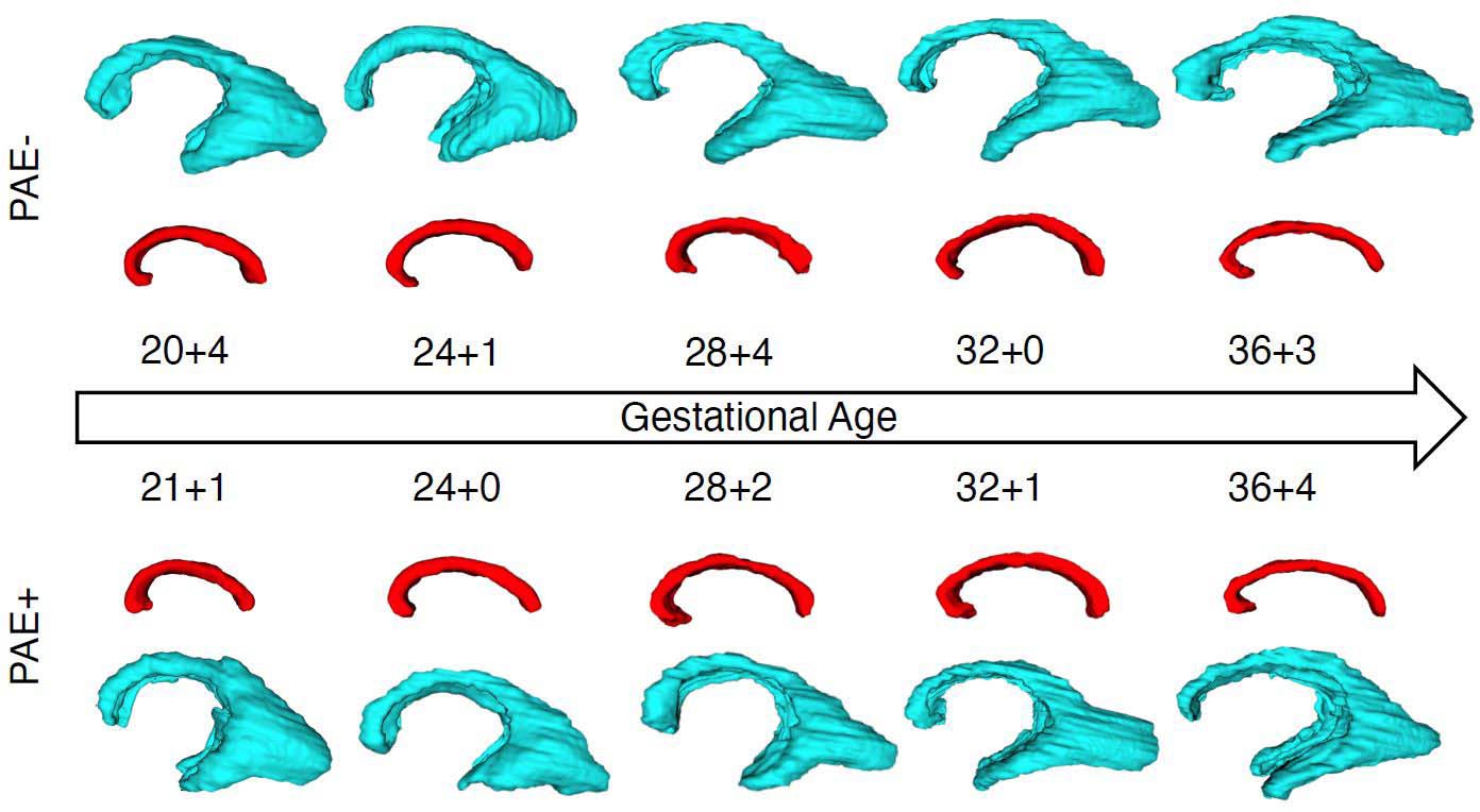
    After eliminating some of the fetal MRIs for reasons such as structural brain anomalies and/or poor image quality, the final study group consisted of 26 fetal MRI exams from 24 alcohol-positive fetuses and a control group of 52 gender- and age-matched healthy fetuses. At the time of imaging, fetuses ranged in age between 20 and 37 weeks.

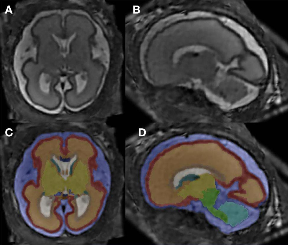
    The researchers used super-resolution imaging, which allowed them to create one dataset to re-construct each fetal brain. Next, they completed an analysis of 12 different brain structures, computing total brain volume and segment volumes of specific brain compartments.

    Longitudinal Growth Trajectories Prenatal Alcohol Exposure
    Longitudinal growth trajectories of the periventricular zone (blue) and the corpus callosum (red) in fetuses with (PAE+) and without (PAE-) prenatal alcohol exposure. The volumetric relations of included images are not representative: The scaling has been altered to allow for an easy comparison of shape and structure of the respective compartments. Credit: RSNA and Marlene Stuempflen, M.D.

    “One of the main hallmarks of our study is that we investigated so many smaller sub-compartments of the brain,” said co-author Marlene Stuempflen, M.D., scientific researcher at the Medical University of Vienna.

    Key Findings: Brain Volume Differences Identified

    The statistical analysis revealed two major differences in the alcohol-exposed fetuses compared to healthy controls: an increased volume in the corpus collosum and a decreased volume in the periventricular zone.

    “This is the first time that a prenatal imaging study has been able to quantify these early alcohol-associated changes,” Dr. Stuempflen said.

    The corpus collosum is the main connection between the brain’s two hemispheres. Dr. Stuempflen noted that it is fitting that this very central structure is affected, because the clinical symptoms of fetal alcohol spectrum disorders are highly heterogenous, or diverse, and cannot be pinpointed to one specific substructure of the brain.

    “The changes found in the periventricular zone, where all neurons are born, also reflect a global effect on brain development and function,” she said.

    Unexpected Patterns in Brain Growth Trajectories

    The researchers said finding a thicker corpus collosum in the alcohol-positive fetuses was surprising because the corpus collosum is thinner in infants with fetal alcohol spectrum disorders.

    “It appears that alcohol exposure during pregnancy puts the brain on a path of development that diverges from a normal trajectory,” Dr. Kasprian said. “Fetal MRI is a very powerful tool to characterize brain development not only in genetic conditions, but also acquired conditions that result from exposure to toxic agents.”

    Additional co-authors are Ernst Schwartz, M.Sc., Mariana Diogo, M.D., Ph.D., Sarah Glatter, M.D., M.M.Sc., Birgit Pfeiler, Victor Schmidbauer, M.D., Lisa Bartha-Doering, Ph.D., Rainer Seidl, M.D., Elisabeth Krampl-Bettelheim, M.D., and Daniela Prayer, M.D.

    Meeting: 107th Scientific Assembly and Annual Meeting of the Radiological Society of North America

    Never miss a breakthrough: Join the SciTechDaily newsletter.
    Follow us on Google and Google News.

    Alcohol Brain Neuroscience Pregnancy Radiological Society of North America
    Share. Facebook Twitter Pinterest LinkedIn Email Reddit

    Related Articles

    How Alcohol Before Birth Alters Brain Wiring and Impacts Behavior for Life

    Does COVID-19 During Pregnancy Harm the Baby’s Brain?

    Study Reveals Wide Spectrum of COVID-19 Brain Complications – Including Stroke, Hemorrhage, and Other Fatal Complications

    Brain’s White Matter Integrity Disrupted in People With Alzheimer’s Gene Mutation

    Right Light on the Mother’s Belly May Be Important to Fetal Brain Development

    Drinking Alcohol Blocks a Chemical in the Brain That Promotes Attention

    Astronaut Brain Volume Changes and Pituitary Gland Deforms During Long Spaceflights

    Alcohol Consumption Is Regulated by Particular Set of Neurons in Specific Brain Region

    Key Brain Region Smaller in Women Taking Birth Control Pills

    Leave A Reply Cancel Reply

    • Facebook
    • Twitter
    • Pinterest
    • YouTube

    Don't Miss a Discovery

    Subscribe for the Latest in Science & Tech!

    Trending News

    Millions of People Have Osteopenia Without Realizing It – Here’s What You Need To Know

    Researchers Discover Boosting a Single Protein Helps the Brain Fight Alzheimer’s

    World-First Study Reveals Human Hearts Can Regenerate After a Heart Attack

    Why Your Dreams Feel So Real Sometimes and So Strange Other Times

    This Simple Home Device May Boost Brain Power in Adults Over 40

    Enormous Prehistoric Insects Puzzle Scientists

    Scientists Develop Bioengineered Chewing Gum That Could Help Fight Oral Cancer

    After 37 Years, the World’s Longest-Running Soil Warming Experiment Uncovers a Startling Climate Secret

    Follow SciTechDaily
    • Facebook
    • Twitter
    • YouTube
    • Pinterest
    • Newsletter
    • RSS
    SciTech News
    • Biology News
    • Chemistry News
    • Earth News
    • Health News
    • Physics News
    • Science News
    • Space News
    • Technology News
    Recent Posts
    • Researchers Identify the Most Common Recessive Neurodevelopmental Disorder Ever Discovered
    • This Is What Makes You Irresistible to Mosquitoes
    • Shockingly Powerful Giant Octopuses Ruled the Seas 100 Million Years Ago
    • After 100 Years, Scientists Uncover Hidden Rule Governing Cosmic Rays
    • The Milky Way Has a Hidden Edge and Scientists Finally Mapped It
    Copyright © 1998 - 2026 SciTechDaily. All Rights Reserved.
    • Science News
    • About
    • Contact
    • Editorial Board
    • Privacy Policy
    • Terms of Use

    Type above and press Enter to search. Press Esc to cancel.