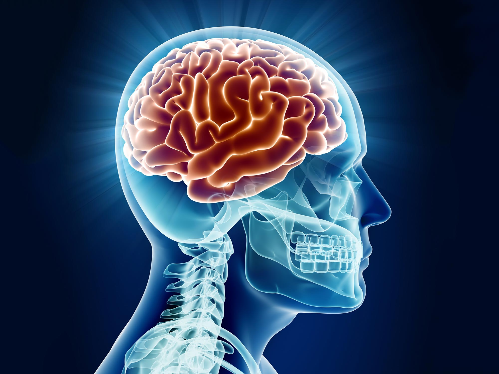
Researchers identify how brain circuits in mice amplify sensory prediction errors through a unique interaction between the neocortex and thalamus, shedding light on mechanisms that may contribute to ASDs and SSDs.
Scientists at the Sainsbury Wellcome Centre at UCL have discovered how the neocortex and thalamus work together to detect discrepancies between what animals expect from their environment and actual events. These prediction errors are implemented by selective boosting of unexpected sensory information. These findings enhance our understanding of predictive processing in the brain and could offer insights into how brain circuits are altered in autism spectrum disorders (ASDs) and schizophrenia spectrum disorders (SSDs).
The study, recently published in Nature, describes how the researchers studied mice in a virtual reality environment to gain insights into the nature of prediction error signals in the brain and the mechanisms behind their development.

Uncovering Neural Mechanisms
“Our brains constantly predict what to expect in the world around us and the consequences of our actions. When these predictions turn out wrong, this causes strong activation of different brain areas, and such prediction error signals are important for helping us learn from our mistakes and update our predictions. But despite their importance, surprisingly little is known about the neural circuit mechanisms responsible for their implementation in the brain,” explained Professor Sonja Hofer, Group Leader at SWC and corresponding author on the paper.
To study how the brain processes expected and unexpected events, the researchers placed mice in a virtual reality environment where they could navigate along a familiar corridor to get to a reward. The virtual environment enabled the team to precisely control visual input and introduce unexpected images on the walls. By using a technique called two-photon calcium imaging, the researchers were able to record the neural activity from many individual neurons in the primary visual cortex, the first area in our neocortex to receive visual information from the eyes.

Enhanced Understanding Through Experiments
“Previous theories proposed that prediction error signals encode how the actual visual input is different from expectations, but surprisingly we found no experimental evidence for this. Instead, we discovered that the brain boosts the responses of neurons that have the strongest preference for unexpected visual input. The error signal we observe is a consequence of this selective amplification of visual information. This implies that our brain detects discrepancies between predictions and actual inputs to make unexpected events more salient” explained Dr Shohei Furutachi, Senior Research Fellow in the Hofer and Mrsic-Flogel labs at SWC and first author on the study.
To understand how the brain generates this amplification of the unexpected sensory input in the visual cortex, the team used a technique called optogenetics to inactivate or activate different groups of neurons. They found two groups of neurons that were important for causing the prediction error signal in the visual cortex: vasoactive intestinal polypeptide (VIP)-expressing inhibitory interneurons in V1 and a thalamic brain region called the pulvinar, which integrates information from many neocortical and subcortical areas and is strongly connected to V1. However, the researchers found that these two groups of neurons interact in a surprising way.
Collaborative Neural Dynamics
“Often in neuroscience we focus on studying one brain region or pathway at a time. But coming from a molecular biology background, I was fascinated by how different molecular pathways synergistically interact to enable flexible and contextual regulation. I decided to test the possibility that cooperation could be occurring at the level of neural circuits, between VIP neurons and the pulvinar,” explained Dr Furutachi.
Indeed, Dr Furutachi’s work revealed that VIP neurons and pulvinar act synergistically together. VIP neurons act like a switchboard: when they are off, the pulvinar suppresses activity in the neocortex, but when VIP neurons are on, the pulvinar can strongly and selectively boost sensory responses in the neocortex. The cooperative interaction of these two pathways thus mediates the sensory prediction error signals in the visual cortex.
Future Research and Implications
The next steps for the team are to explore how and where in the brain the animals’ predictions are compared with the actual sensory input to compute sensory prediction errors and how prediction error signals drive learning. They are also exploring how their findings could help contribute to understanding ASDs and SSDs.
“It has been proposed that ASDs and SSDs both can be explained by an imbalance in the prediction error system. We are now trying to apply our discovery to ASDs and SSDs model animals to study the mechanistic neural circuit underpinnings of these disorders,” explained Dr Furutachi.
Reference: “Cooperative thalamocortical circuit mechanism for sensory prediction errors” by Shohei Furutachi, Alexis D. Franklin, Andreea M. Aldea, Thomas D. Mrsic-Flogel and Sonja B. Hofer, 28 August 2024, Nature.
DOI: 10.1038/s41586-024-07851-w
This research was funded by the Sainsbury Wellcome Centre Core Grant from the Gatsby Charity Foundation and Wellcome (219627/Z/19/Z and 090843/F/09/Z); a Wellcome Investigator Award (219561/Z/19/Z); the Gatsby Charitable Foundation (GAT3212 and GAT3361); the Wellcome Trust (090843/E/09/Z and 217211/Z/19/Z); European Research Council (HigherVision 337797; NeuroV1sion 616509); the SNSF (31003A 169525); Biozentrum core funds (University of Basel).
Never miss a breakthrough: Join the SciTechDaily newsletter.
Follow us on Google and Google News.
3 Comments
Yeah, that mechanism is called “thinking”.
It’s how a tortured mouse responds to torture, “head-fixed, food-deprived mice running on a cylinder”.
According to the actual study, “To study the neural implementation of predictive processing in cortical sensory networks, we used a paradigm in which head-fixed, food-deprived mice running on a cylinder navigated a virtual corridor in which they developed spatial predictions about stimulus identity at particular locations along the corridor.”
Only psychopaths would do this. “head-fixed, food-deprived mice running on a cylinder”. Sick. This is torture. No relevance to anything other than the psychopathology of the researchers.
This is why I quit medicine. It is saturated with sick research like this. See my article, Animal Research: The Rot at the Core of Medicine. https://www.academia.edu/123055005/Animal_Research_The_Rot_at_the_Core_of_Medicine