New pathway reveals how misfolded proteins are cleared from the nucleus, offering insights for neurodegenerative disease treatments.
Misfolded proteins pose a threat to cellular health, as they interfere with normal functions and contribute to age-associated degenerative conditions such as Alzheimer’s, Parkinson’s, and Huntington’s diseases. The mechanisms by which cells eliminate these harmful proteins are not yet fully understood.
A recent study, published on April 20 in Nature Cell Biology, reveals groundbreaking findings by Stanford University researchers. They uncovered a previously unidentified cellular pathway that facilitates the removal of misfolded proteins from the nucleus, where the cell’s DNA is stored, transcribed, and replicated. Maintaining the integrity of these processes is crucial for proper cellular function. This newly discovered pathway offers potential therapeutic targets for treating age-related diseases.
To find the new pathway, researchers in the lab of Judith Frydman, the Donald Kennedy Chair in the School of Humanities and Sciences, integrated several genetic, imaging, and biochemical approaches to understand how yeast cells dealt with misfolded proteins. For the experiments, the team restricted misfolded proteins to either the nucleus or the cytoplasm – the area inside the cell but outside the nucleus. The team visually followed the fate of the misfolded proteins through live-cell imaging and super-resolution microscopy.
“The first exciting thing was that we actually found that there’s communication between the nucleus and the cytoplasm,” said Emily Sontag, the co-lead author of the paper and a former postdoctoral student in the Frydman Lab. “So they’re telling each other, ‘We both have a lot of misfolded proteins; let’s coordinate to send them here to this garbage dump so that they can be removed.’”
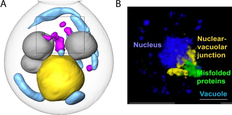
The team identified the “garbage dump” site as the intersection of the nucleus and the vacuole – an organelle full of enzymes for degrading proteins – and showed that misfolded proteins in this “garbage dump” site are moved into the inside of the vacuole for degradation. They also showed that the pathway depends on a class of proteins used to create small vesicles for transporting molecules around cells.
“Tying that particular family of proteins and this aspect of vesicle traffic biology to protein clearance gives us a new way to look at Alzheimer’s, Parkinson’s, Huntington’s – all these neurodegenerative diseases,” said Sontag.
Shared ‘Garbage Dump’ Site for the Nucleus and the Cytoplasm
Cells can deal with misfolded proteins in two ways: by refolding them or by eliminating them. A third option is to store them at a specific cellular location.
“While the cell decides whether to refold or degrade proteins, it sequesters them into these membrane-less inclusions,” said Frydman, who is senior author of the paper. Inclusions are clusters of misfolded proteins that occur in both the cytoplasm and in the nucleus.
The team found that the cellular machinery forms small misfolded-protein inclusions in different places within the nucleus and cytoplasm, like tiny garbage dumps, that then migrate toward the boundary between the nucleus and the vacuole, a bigger garbage dump. Eventually, the nuclear and cytoplasmic misfolded protein inclusions line up to face each other, with the nuclear envelope separating them.
“The communication back and forth between the nucleus and the cytoplasm was not something we expected at all,” said Sontag. “Knowing that those two compartments can kind of work together to clear garbage from everywhere was really awesome.”
“It shows that the management of misfolded proteins in the nucleus and the management of misfolded proteins in the cytoplasm are distinct but are coordinated,” said Frydman. “And what is really cool is that each compartment moves their misfolded proteins to the site where the nuclear envelope meets the vacuolar membrane.”
From Dump Site to Degradation – a New Pathway
The vacuole in yeast is equivalent to the lysosome in mammalian cells. It’s a membrane-bound organelle filled with enzymes that break down proteins – a recycling center for the cell.
“This is not random,” said Fabián Morales-Polanco, the co-lead author of the paper and a postdoctoral scholar in the Frydman lab. “The cell is bringing inclusions to the same spot for a reason.”
The team suspected that reason was to send the inclusions to the vacuole for degradation, but that raised further questions. It’s easy for cytoplasmic inclusions to enter the vacuole by autophagy – a process cells use to pull things from the cytoplasm into the vacuole or lysosome. But in the nucleus, inclusions are separated from the vacuole by the nuclear envelope.
“Even though they come to the same spot, they don’t get into the vacuole by the same door,” said Morales-Polanco.
To investigate the pathways of damaged proteins into the vacuole, the team blocked the proteasome – the other major protein clearance mechanism – and monitored the remaining protein clearance activity. They also created 3D images of the cells containing these misfolded protein inclusions using cryogenic soft X-ray tomography and fluorescence microscopy data.
They found that the cytoplasmic inclusions did push into the vacuole, as expected. But the route for the nuclear inclusions was surprising. The nuclear inclusions budded straight from the nucleus into the vacuole at the junction of the two membranes. Using a series of genetic experiments, the team showed that ESCRT II/III and Vps4 proteins facilitated that budding-into-the-vacuole action. These proteins are known to cause membranes to bend and “bud,” or form new vesicles in other processes, but have not been studied as helping clear the nucleus of damaged proteins. They may be attractive therapy targets for misfolded protein diseases.
Finally, using pH-sensitive tags, the team actually followed inclusions into the vacuole.
“We were able to see these misfolded proteins entering into the vacuole and show this is really a new pathway,” said Morales-Polanco.
An Eye on Aging
The team did these experiments in yeast cells, which are easy to grow and quick to reproduce. One next step is to investigate whether this same pathway is used in mammalian cells to clear human disease-related proteins.
Another next step is to define how the communication between the nucleus and cytosol happens along the pathway, and yet another is to see how the pathway is affected by aging.
“There’s a lot of evidence that this process for dealing with misfolded proteins slows down with age,” said Sontag. “So, as time goes on, aged cells are not able to remove all that garbage as quickly or as efficiently, and misfolded proteins build up more and more inside the cell.”
“We showed that nuclear and cytoplasmic quality control pathways communicate via the nuclear envelope, a structure that is impaired by aging and by neurodegenerative disease,” said Frydman. “Many progeria mutants, which cause premature aging, distort the nuclear envelope. This work really is a game changer in finally bringing a new way to understand, and hence cure, a wide range of terrible diseases that affect an increasingly aged population.”
Reference: “Nuclear and cytoplasmic spatial protein quality control is coordinated by nuclear–vacuolar junctions and perinuclear ESCRT” by Emily M. Sontag, Fabián Morales-Polanco, Jian-Hua Chen, Gerry McDermott, Patrick T. Dolan, Daniel Gestaut, Mark A. Le Gros, Carolyn Larabell and Judith Frydman, 20 April 2023, Nature Cell Biology.
DOI: 10.1038/s41556-023-01128-6
The study was funded by the National Institutes of Health, Way Klingler Faculty Development Awards from Marquette University, Pew Charitable Trusts, and the Gordon and Betty Moore Foundation.
Never miss a breakthrough: Join the SciTechDaily newsletter.
Follow us on Google and Google News.