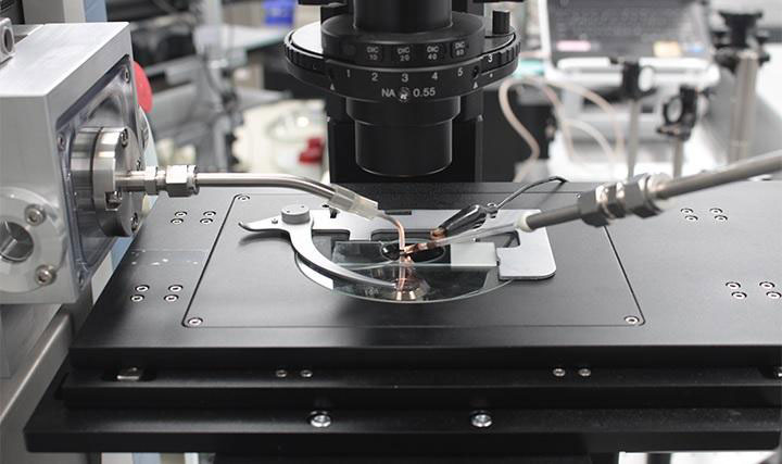
High resolution atmospheric pressure mass spectrometry imaging system. Credit: Daegu Gyeongbuk, Institute of Science and Technology (DGIST)
A team of researchers at DGIST has recently developed a technology which enables to acquire a high-resolution mass spectrometry imaging in micrometer size of live biological samples without chemical pretreatment in the general atmospheric pressure environment. The newly developed mass spectrometry imaging system to analyze living biological samples in the atmospheric pressure environment at a resolution of 3 μm and could be applied in molecular biology and medical diagnosis fields.
This achievement has been led by Professor Dae Won Moon and Dr. Jae Young Kim from the department of New Biology at DGIST. The study demonstrates a high-resolution mass spectrometry imaging system capable of analyzing live biological samples at a resolution of several micrometers (㎛).
Mass spectrometry imaging system is a technology to measure how much of a substance exists in a certain region as it acquires biomolecular information of tissues and cells as well as the spatial distribution of biomolecules through the measurement of the mass of biomolecules by desorbing biomolecules from tissues and cells.
Researchers typically use an ion beam desorption system or a laser desorption method, in which biomolecule samples are separated in a vacuum state, to obtain a high-resolution mass spectrometric image. However, in order to accurately analyze the sample by placing it in a vacuum chamber, pretreatment processes such as cutting the frozen samples or chemical treatment were required. In the process, side effects such as damaging the samples or loss of molecular information occurred.
Although researches on mass spectrometry and mass spectrometry imaging methods in the atmospheric pressure environment have been carried out around the world over the last 10 years, they have not been directly applied in biomedical science and medical areas due to the performance limitation of ionizing biological samples under atmospheric pressure.
In the study, the research team used a femtosecond laser to desorb biomolecules from biological samples and a plasma jet to ionizing biomolecules and analyzed mass spectrometry of biological samples at the same time. Furthermore, the researchers equally spread gold nanoparticles on a biological sample by utilizing the endocytosis of live tissues, and changed the light absorption properties of biological samples so that biomolecule desorption can easily occur with low laser power.
In order to solve engineering problems that may occur during atmospheric pressure ionization mass spectrometry of biological samples, they added ion transmission device, a laser focusing lens, a 2D scanning stage, and a signal synchronization circuit between devices and completed the system.
Using this system, about 250 biomolecule substances were extracted from hippocampal tissue sections of mouse brain, and mass spectrometry imaging with a resolution of 3 μm or less was obtained from 10 biomolecule materials. In addition, adjacent tissue sections taken from the same rats were used to determine the effectiveness of the drug at the biopsy level.
Through the findings of this study, it is expected that the reliability of new drug development can be improved and the sacrifice of laboratory animals can be reduced by using a mass spectrometry imaging system as an organization-based drug screening technology.
Professor Moon stated, “You can acquire a large amount of undamaged biomolecule information from biological samples that have metabolic activity. At the same time, you can visualize it in high resolution. Therefore, this technology will significantly contribute to molecular biology research.” He also added “We will carry out further studies to widen the molecular weight range that are detectable in the sample and utilize them in the field of medical diagnoses such as the development of new drug screening and mass spectrometric endoscopy.”
Reference: “Atmospheric pressure mass spectrometric imaging of live hippocampal tissue slices with subcellular spatial resolution” by Jae Young Kim, Eun Seok Seo, Hyunmin Kim, Ji-Won Park, Dong-Kwon Lim and Dae Won Moon, 13 December 2017, Nature Communications.
DOI: 10.1038/s41467-017-02216-6
This study was published in the online edition of ‘Nature Communications,’ sister journal of the international journal Nature, on December 13. The study was collaborated with the research team of Professor Dong-Kwon Lim of Korea University and the research team of Professor Ji-Won Park of Chungnam University.






Be the first to comment on "New Imaging Technique Makes Diagnosis Easier and Smarter"