
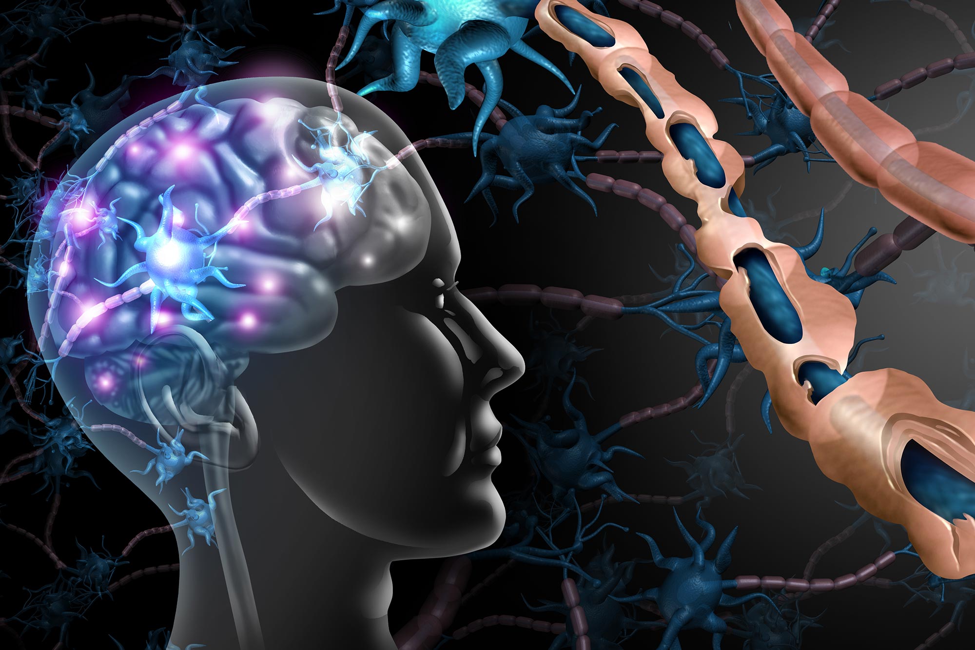
Researchers have developed an artificial intelligence method that can uncover previously hidden signs of multiple sclerosis in routine MRI scans.
For decades, multiple sclerosis specialists have been forced to study the disease through an incomplete picture. Standard magnetic resonance imaging (MRI) can reveal damage in the brain’s white matter, but some of the lesions most closely tied to disability and cognitive decline have remained largely invisible.
Those hidden injuries occur in the gray matter, particularly the brain’s cortex. Researchers have long known that cortical lesions can offer important clues about how MS progresses and how severely it may affect a patient. Yet routine MRI has not provided a reliable way to detect or monitor them, leaving clinicians with a major blind spot. Even many of the newer drugs that can slow MS progression are designed primarily around reducing white matter lesions.
Now, a University at Buffalo-led research team has used artificial intelligence to expose some of that previously concealed damage in existing MRI scans. Published in Communications Medicine, the study shows how computational methods can compare information across multiple images and recover disease signals that conventional viewing methods fail to capture.
“Detecting previously invisible cortical lesions on conventional legacy MRI scans has major implications for MS research and clinical care,” says Robert Zivadinov, MD, PhD, senior author on the paper, SUNY Distinguished Professor in the Department of Neurology and director of the Buffalo Neuroimaging Analysis Center (BNAC) in the Jacobs School of Medicine and Biomedical Sciences at UB. “The ability to see for the first time these previously hidden indicators of MS disease progression, including cognitive impairment and disability, is an important advance,” he says.
Hidden lesions shaped MS uncertainty
Cortical lesions have been linked to MS since nearly the time the disease was first described in the late 19th century. Still, they did not become part of diagnostic criteria until the 21st century, and even then, their usefulness was limited because clinical MRI could not reliably reveal them.
“We have all been very frustrated, knowing that these cortical lesions were there but not being able to see them,” says Michael G. Dwyer, PhD, first and corresponding author on the paper, associate professor of neurology and biomedical informatics in the Jacobs School and a researcher with BNAC. “There’s a lot of ongoing damage that continues to happen in MS that you won’t see with conventional MRI, but that histopathologists have been clearly demonstrating for decades on postmortem tissue.
“What this collaboration has been able to accomplish is a real success story for applying AI in the medical arena,” he continues. “We now have access to these incredibly useful data on MRI scans that were there but you couldn’t see them without using AI to pull them out. The computational methods are finally at the point where we can do this.”
The researchers used AI methods that build on work by coauthors in the Netherlands. The goal was to recover information that is not visible in any one image alone, but becomes detectable when the relationships among several images are analyzed together.
To do that, they combined multiple image processing methods, including a new approach called MMCLE, or multimodal cortical lesion enhancement. They then tested the methods on MRI scans from ORATORIO, a large phase III FDA regulatory clinical trial of the MS drug Ocrelizumab that included more than 700 participants.
More than 11,000 cortical lesions detected
On standard scans, individual brain images mostly showed white matter lesions. After the researchers applied AI-guided image processing across different contrast images, a hidden layer of damage became visible. The method detected about 15 to 20 cortical lesions per patient, adding up to more than 11,000 lesions across the full dataset.
“If you look on the original scans, you generally can’t see the cortical lesions,” says Dwyer, “but generative AI is very powerful because it can look between the scans and detect tiny differences between them. Because it sees those minor discrepancies, AI can reveal that there’s something going wrong there, that the tissue is not behaving like healthy tissue. The trained models can view multiple MRI images together and synthesize them, and synthesize what had been missing.”
The international project was led by UB and included scientists and clinicians from academic institutions and industry, including Genentech, which makes Ocrelizumab. Zivadinov says that the range of expertise involved was central to the result.
“This work, which has revealed that there is so much invisible pathology in the brain, will have tremendous impact for reviewing data from past clinical trials and also for those going forward,” he says.
Reference: “Quantifying cortical lesions in multiple sclerosis MRI datasets using multi-contrast post-processing and deep learning” by Michael G. Dwyer, Niels Bergsland, Alexander Bartnik, Dejan Jakimovski, Samantha Noteboom, Menno M. Schoonheim, Martijn D. Steenwijk, Jinglan Pei, David Clayton and Robert Zivadinov, 7 July 2026, Communications Medicine.
DOI: 10.1038/s43856-026-01683-7
This work was supported in part by Genentech, Inc. as part of a research collaboration.
Never miss a breakthrough: Join the SciTechDaily newsletter.
Follow us on Google and Google News.