Research led by UCSF has pinpointed a hallmark of disease repair that could be utilized in the development of future therapies.
Ten years following the discovery of a common antihistamine, clemastine, as a potential treatment for multiple sclerosis by scientists at UC San Francisco, a new method to evaluate the drug’s efficacy in repairing the brain has been developed. This development could also facilitate the assessment of potential future treatments for the devastating disorder.
Under the leadership of physician-scientist Ari Green, MD, who alongside neuroscientist Jonah Chan, Ph.D., initially pinpointed clemastine’s potential therapeutic role for MS, MRI scans were used to investigate the drug’s effects on the brains of 50 clinical study participants.
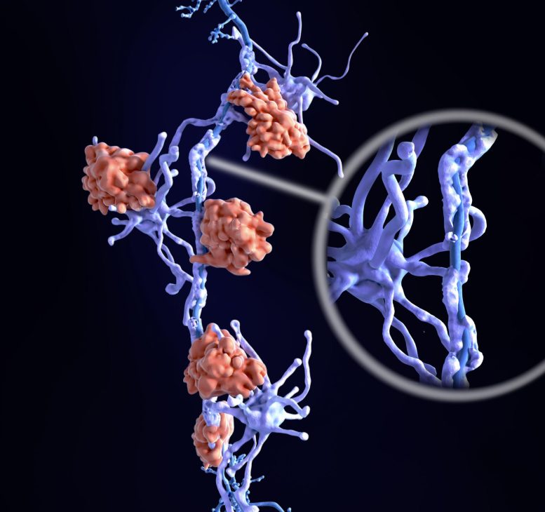
In MS, patients lose myelin, the protective insulation around nerve fibers. This myelin loss triggers delays in nerve signals, leading to weakness and spasticity, vision loss, cognitive slowing, and other symptoms.
In the brain, water trapped between the thin layers of myelin that wrap nerve fibers cannot move as freely as water floating between brain cells. This unique property of myelin allowed imaging experts to develop a technique to compare the difference in myelin levels before and after the drug was administered, by measuring the so-called myelin water fraction, or the ratio of myelin water to the total water content in brain tissue.
In their study, published May 8, 2023, in PNAS, the researchers found that patients with MS who were treated with clemastine experienced modest increases in myelin water, indicating myelin repair. They also proved that the myelin water fraction technique, when focused on the right parts of the brain, could be used to track myelin recovery.
“This is the first example of brain repair being documented on MRI for a chronic neurological condition,” said Green, medical director of the UCSF Multiple Sclerosis and Neuroinflammation Center and a member of the Weill Institute for Neurosciences. “The study provides the first direct, biologically validated, imaging-based evidence of myelin repair induced by clemastine. This will set the standard for future research into remyelinating therapies.”
Myelin Increased Even After Medication Was Stopped
In the study, patients with MS who enrolled in the ReBUILD trial were divided into two groups: the first group received clemastine for the first three months of the study and the second group received clemastine only in months three to five. Using the myelin water fraction as a biomarker, the researchers found that myelin water increased in the first group after participants received the drug and continued to increase after clemastine was stopped. In the second group, the myelin water fraction showed decreases in myelin water in the first portion of the study, under the placebo, and a rebound after participants received clemastine.
The findings corroborate the results of a previous study with the same 50 patients that had found the allergy medication reduced delayed nerve signaling, potentially alleviating symptoms.
In the current study, researchers looked at the corpus callosum, a region of the brain with a high myelin content that connects the right and left hemispheres. They found that significant repair occurred outside the visible lesions typically associated with MS. This underscores the need to focus on myelin repair beyond these lesion sites.
Clemastine works in this setting by stimulating the differentiation of myelin-making stem cells. This places the medication a generation ahead of existing MS drugs that work by dampening the activity of the immune system, calming inflammation, and reducing the risk of relapse. It still isn’t ideal, though, making the water fraction measurement an important tool in developing better therapeutics.
“Clemastine can only be partially effective at the doses we can use,” said Green, who is also a neuro-ophthalmologist and chief of the Division of Neuroimmunology and Glial Biology in the UCSF Department of Neurology. “It can be sedating, which may be especially undesirable in patients with MS. We are hopeful better medications will be developed, but clemastine has proven to be the tool to show remyelination is possible.”
Proposed future research will examine clemastine’s potential in treating brain injury in premature infants, who often experience myelin damage. Pediatric neurologist Bridget Ostrem, MD, Ph.D., of UCSF Benioff Children’s Hospitals, is currently seeking approval from the Food and Drug Administration to initiate the first clinical trial testing clemastine to treat this debilitating and disabling condition.
Reference: “MWF of the corpus callosum is a robust measure of remyelination: Results from the ReBUILD trial” by Eduardo Caverzasi, Nico Papinutto, Christian Cordano, Gina Kirkish, Tristan J. Gundel, Alyssa Zhu, Amit Vijay Akula, W. John Boscardin, Heiko Neeb, Roland G. Henry, Jonah R. Chan and Ari J. Green, 8 May 2023, Proceedings of the National Academy of Sciences.
DOI: 10.1073/pnas.2217635120
The study was supported by The Rachleff Family Westridge Foundation, Janet Lustgarten and the Lustgarten Family Whitney Fund, and the Adelson Medical Research Foundation.
Never miss a breakthrough: Join the SciTechDaily newsletter.
Follow us on Google and Google News.