
Scientists have developed a new approach to X-ray imaging that captures significantly more light without sacrificing image sharpness.
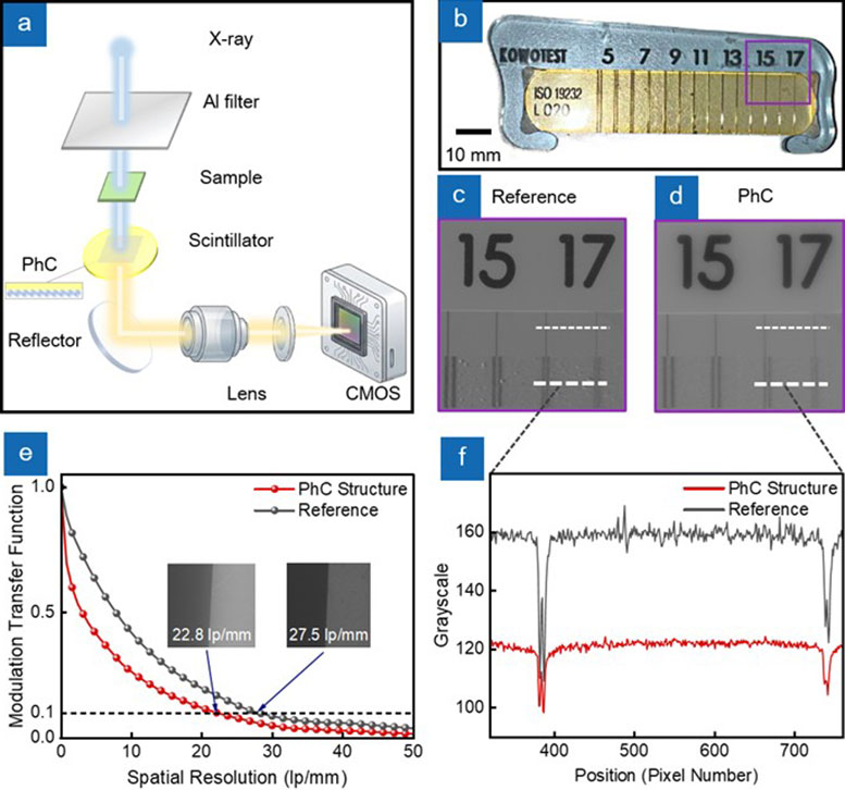
A major obstacle to sharper X-ray images is that much of the light generated inside detectors never reaches the camera. Researchers have developed a nanostructured surface that increased average light output by 4.64 times and improved the imaging signal-to-noise ratio by 13 decibels. The experimental system also maintained spatial resolution finer than 25 μm (about 0.001 inch).
Developed by scientists at Chongqing University, the 26th Institute of CETC, and Stevens Institute of Technology, the approach could help X-ray detectors produce clearer images when very few photons are available. Potential applications include medical scans using lower radiation doses, security screening, and detailed industrial inspections.
Why X-Ray Detectors Trap Their Own Light
X-ray imaging relies on materials called scintillators to convert invisible X-ray radiation into visible light. These “photosensitive” components act as a “translator,” converting the “language” of high-energy radiation into optical signals that cameras or photodetectors can capture and turn into images.
High-density inorganic scintillators (such as GAGG) are particularly effective at absorbing X-rays and generating light, but much of that light cannot escape. Because these materials have a substantially higher refractive index than their surroundings, light striking the surface at certain angles undergoes total internal reflection. Up to 90% of the generated photons can become trapped in guided optical modes, reducing the amount of light available for imaging.
This problem becomes more severe when detectors operate with weak X-ray signals. Increasing scintillator thickness can improve X-ray absorption, but it also allows light to spread sideways before detection, reducing image sharpness. Thinner scintillators preserve fine details but can produce weaker signals.
Using Photonic Crystals to Release Trapped Light
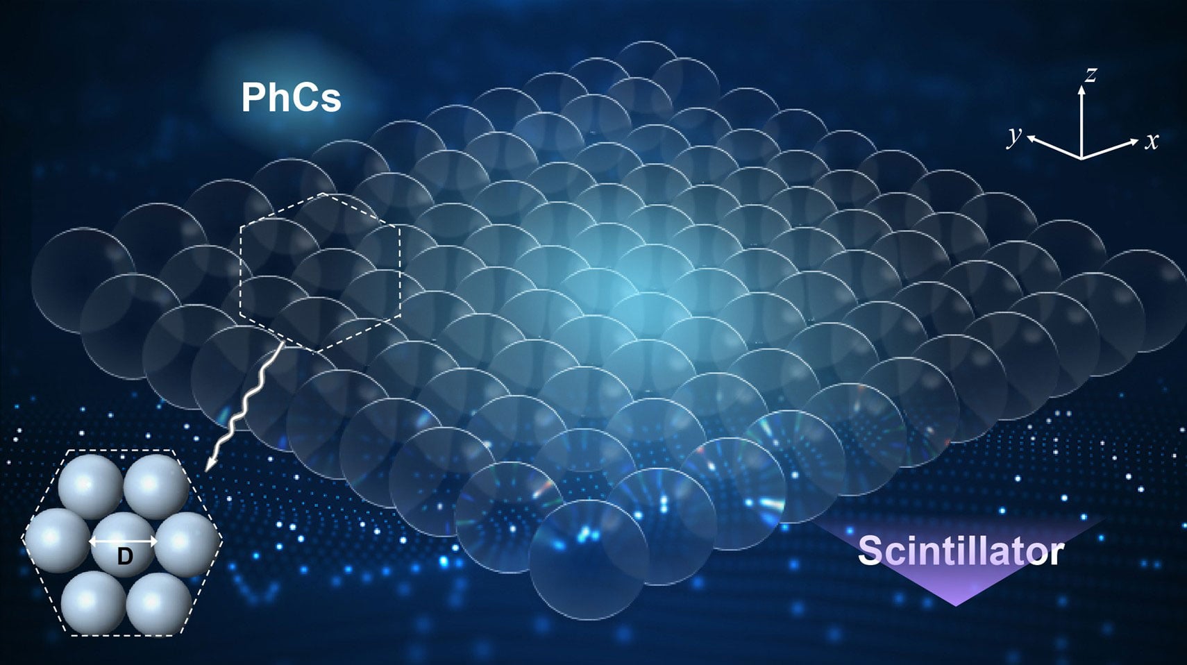
The researchers addressed this trade-off by modifying how light escapes the scintillator rather than increasing its thickness. Their design uses a photonic crystal, a regularly repeating nanoscale structure that changes how light travels through a material.
Through a process known as Bragg diffraction, the periodic structure provides additional momentum that allows previously trapped light to escape into free space. This increases the amount of visible light reaching the detector without requiring a thicker scintillator.
Manufacturing such structures over large areas has been difficult. Conventional lithography offers precise patterning but requires costly equipment and complicated processing, particularly on brittle scintillator surfaces. Simpler self-assembly techniques can cover larger areas, but cracks, missing particles, and poorly aligned crystal regions can reduce light extraction and introduce imaging artifacts.
A New Manufacturing Method Driven by Surface Tension
The team developed a self-assembly technique that uses the Marangoni effect, in which differences in surface tension drive liquid movement. By combining ethanol and water, the researchers created surface tension gradients that helped organize polystyrene nanospheres into a highly ordered layer.
Surfactants helped pack the particles more tightly, while a vibration-free drainage process transferred the assembled structure onto a 25 mm (about 0.98 inch) diameter GAGG scintillator wafer. The resulting photonic crystal consisted of a continuous monolayer of nanospheres arranged in a hexagonal close-packed pattern.
The research was led by Associate Professor Feng Peng of Chongqing University, working with Dr. Ding Yuchong of the 26th Institute of CETC and PhD candidate Kaifeng Liu of Stevens Institute of Technology. Their paper was published in Opto-Electronic Advances.
Toward Clearer Images With Less Radiation
The experimental findings demonstrate that modifying the scintillator surface can substantially improve light extraction while preserving fine image details. This approach could be particularly useful in photon-starved conditions, where limited X-ray exposure makes it difficult to distinguish weak signals from background noise.
Further development is still needed before the technology can be widely adopted. Residual structural defects can affect spatial resolution, and the researchers plan to refine the assembly process and investigate compatibility with other scintillator materials.
This study was financially supported by the Natural Science Foundation of Chongqing, China under grant CSTB2025NSCQ-LZX0041, the Development Foundation of 26th Institute of China Electronics Technology Group Corporation under grant K2605ZZ2025002, the Beijing Natural Science Foundation under Grant L242114 and the National Key Research and Development Program of China under Grant 2019YFC0605203.
Never miss a breakthrough: Join the SciTechDaily newsletter.
Follow us on Google and Google News.