Researchers have captured the first atomic-level views of human SMUG1, a key enzyme involved in repairing damaged DNA.
Every cell in your body is constantly battling damage to its DNA. Now, scientists have captured the first atomic level images of a key human repair enzyme in action, revealing how cells identify and remove some of the molecular mistakes that can threaten genetic stability.
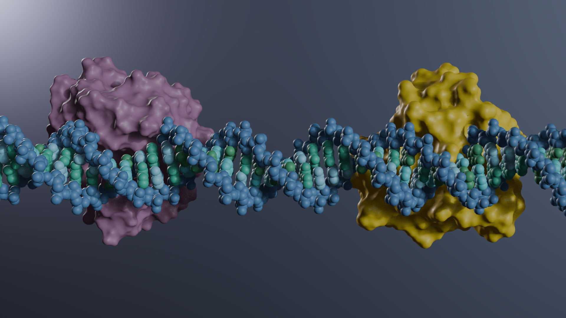
The enzyme, known as SMUG1, serves as part of the cell’s quality control system. Researchers have now mapped its structure in unprecedented detail, providing new insight into how it locates and removes damaged DNA components. The findings could also help guide future efforts to develop drugs that target this important repair pathway.
DNA is constantly damaged by normal processes in our cells, as well as by environmental factors and cancer treatments. If the damage is not repaired, it can lead to permanent mutations. One enzyme involved in this repair process is human SMUG1, which removes uracil and related damaged bases from DNA. Uracil is one of the four nucleotide bases normally found in the nucleic acid RNA, but when it appears in DNA, it can cause problems if not repaired.
Although SMUG1 has long been known to play an important role in DNA repair, its three-dimensional structure had remained unknown. In the new study, published in Nature Communications, researchers captured the enzyme in several forms, including alone, bound to uracil and 5-fluorouracil, and attached to double-stranded DNA.
Linked to cancer biology
5-fluorouracil is a chemotherapy drug widely used to treat cancer. When the compound becomes incorporated into DNA, SMUG1 helps remove it. Because the enzyme is involved in both DNA repair and cancer biology, the newly determined structures could provide a starting point for designing drugs that specifically target SMUG1.
The research also produced the first combined neutron and X-ray structure of a DNA-binding protein.
“This provides rare insight into proton positions and hydrogen-bonding networks in the enzyme active site, details that are often difficult to resolve with X-ray crystallography alone,” Stenmark said.
The project involved researchers from Uppsala University, Karolinska Institutet, Institut Laue-Langevin (ILL), the European Spallation Source (ESS), and Stockholm University.
“The finding is especially timely for Sweden, as the European Spallation Source (ESS), the world’s most powerful neutron source, is currently being built in Sweden; this will dramatically expand opportunities for studies of this kind,” Stenmark said.
Taken together, the findings provide a detailed look at how SMUG1 repairs damaged DNA and offer a valuable foundation for future efforts to develop therapies that target this important repair mechanism.
Reference: “Structural basis for uracil removal from DNA by human SMUG1” by Julian M. Ludäscher, Emma Scaletti Hutchinson, Guillem Vila-Julià, Ann-Sofie Jemth, Saher Shahid, Elisee Wiita, Israel Cabeza de Vaca, Szymon Pach, Lukas Gajdos, Swati Aggarwal, Ellen Walse, Oliver Mortusewicz, Thomas Helleday, Jens Carlsson and Pål Stenmark, 2 June 2026, Nature Communications.
DOI: 10.1038/s41467-026-72937-0
Funding: Swedish Cancer Foundation, Vetenskapsrådet, Barncancerfonden, EU-IMI2, Wallenberg Scholars, Cancer Research Funds of Radiumhemmet, the Marie Skłodowska- Curie PRISMAS program.
Never miss a breakthrough: Join the SciTechDaily newsletter.
Follow us on Google and Google News.