
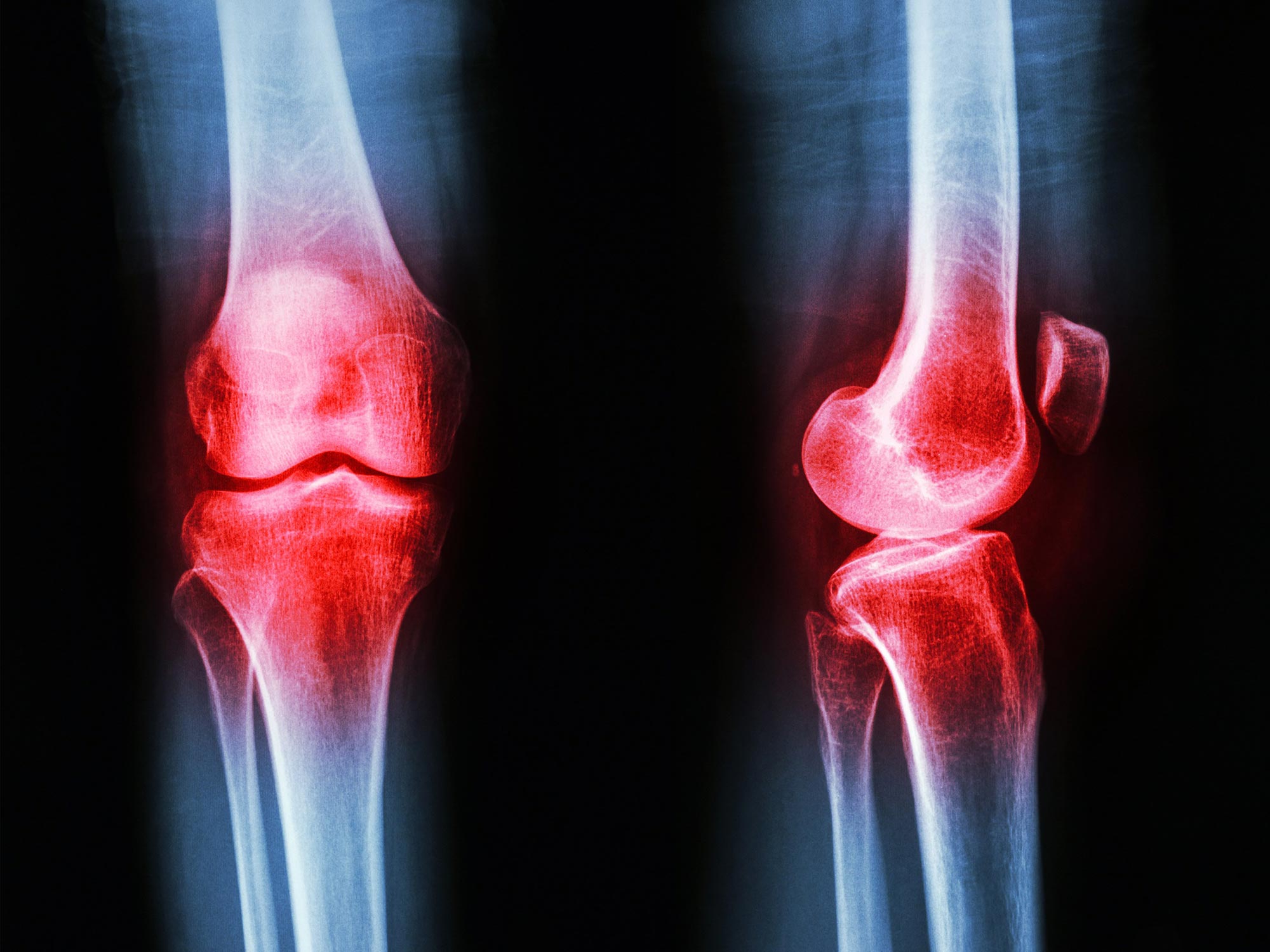
Some joints may be predisposed to rheumatoid arthritis by cellular and structural differences established before birth.
Some joints may be predisposed to inflammatory arthritis before a person is even born. Researchers at the Kennedy Institute have identified developmental differences that could help explain a longstanding puzzle in rheumatoid arthritis: why the disease repeatedly affects certain joints while leaving others relatively untouched.
Their study, published in Nature Immunology, examines whether differences built into joint tissues during development contribute to this selective pattern.
Rheumatoid arthritis is an autoimmune disease in which the immune system attacks the synovium, the tissue lining a joint, causing pain, swelling, and stiffness. Persistent inflammation can eventually damage cartilage, bone, and surrounding tissues.
Vulnerability appears before birth
To look for an explanation, the researchers compared two types of finger joints with very different susceptibility to rheumatoid arthritis. Proximal interphalangeal (PIP) joints are commonly affected, while distal interphalangeal (DIP) joints near the fingertips are usually spared.
The PIP joints had a larger volume of synovial tissue and contained higher levels of PI16-positive (PI16+) fibroblasts, specialized connective tissue cells. Because these differences were already present before birth, the findings suggest that the tissues of individual joints may help determine their later susceptibility to disease.
Christopher Buckley, Kennedy Professor of Translational Rheumatology at the University of Oxford, said: “For decades we have known that rheumatoid arthritis selectively targets particular joints, but one of the great unanswered questions is why? Our findings suggest that the answer lies not only in the immune system but also in the tissues themselves. The cellular and structural characteristics established during development may help determine where inflammation takes hold later in life.”
Developing joints reveal their differences
The researchers mapped developing human finger joints using single-cell sequencing, advanced image analysis, and high-resolution 3D X-ray scanning. Examining joints during development allowed them to study entire joints in a level of detail that is difficult to achieve with adult tissue.
The developing joints consisted mainly of structural cells, including cartilage-forming cells and fibroblasts, rather than immune cells. The researchers then explored the signals that guide these cells toward their different specialized forms.
Synovial lining fibroblasts emerged as one population of particular interest. These cells make substances that lubricate joints and support smooth movement, but they can also behave abnormally in arthritis. The analysis indicated that the synovial lining may develop from two sources, cartilage cells and fibroblasts surrounding the joint. Localized conditions, including low oxygen levels, appeared to influence this process. Understanding those signals could provide insight into how these cells function and potentially how their normal protective role might be restored in disease.
Vulnerable joints contain distinct fibroblasts
Further comparisons exposed clear differences between PIP and DIP joints. Using a custom image analysis tool, the researchers found that PI16+ fibroblasts were more abundant in PIP joints and concentrated around blood vessels and where tendons and ligaments connect with surrounding tissue.
These cells also reacted differently to inflammatory signals than other fibroblast populations. Although PI16+ and PI16- fibroblasts shared a common pro-inflammatory response, PI16+ cells showed additional changes in pathways involved in tissue organization and immune regulation.
Structural differences were also apparent. High-resolution 3D imaging at Diamond Light Source on the Harwell Science and Innovation Campus showed that the synovial tissue surrounding PIP joints was larger and organized differently from the tissue in joints that rheumatoid arthritis usually spares. Taken together, these cellular and structural features may help explain why inflammation becomes established in some joints but not others.
Dr. Sarah Davidson, Postdoctoral Researcher at the Kennedy Institute and one of the first authors of the study, said: “We found that joints commonly affected by rheumatoid arthritis already contain distinct cellular populations before birth. PI16+ fibroblasts were enriched in vulnerable joints and responded differently to inflammatory signals. Their location and behavior suggest they could help shape where disease develops.”
The findings suggest that the characteristic pattern of rheumatoid arthritis may partly reflect joint architecture established during development. Rather than depending on immune activity alone, susceptibility to inflammation may also be influenced by the particular cellular and structural environment present within each joint.
Reference: “The embryonic origins of site-specific arthritis” by Sarah Davidson, Davide Simone, Kathrin Jansen, Max Cowan, Caio Machado, Ian Reekie, Ananya Bhalla, Rowie Borst, Cesar Prada Medina, Joshua Bull, Zhi Yi Wong, Sarah Hill, Micon Garvilles, Sam Pledger, Patricia Reis Nisa, Nora Rebecca Schwingen, Dylan Windell, Moustafa Attar, Catherine Disney, Andrew J. Bodey, Alissa Parmenter, Helen Byrne, Sharif Ahmed, Shashidhara Marathe, Peter D. Lee, Chris Mahony, Adam P. Croft, Stephen Sansom, Mark C. Coles and Christopher D. Buckley, 8 June 2026, Nature Immunology.
DOI: 10.1038/s41590-026-02542-2
Supported by the Medical Research Council (MRC).
Never miss a breakthrough: Join the SciTechDaily newsletter.
Follow us on Google and Google News.